Goparaju Balaji, Rana Kunjan D, Calabro Finnegan J, Vaina Lucia Maria
Department of BME, Boston University, Boston, USA.
Med Sci Monit. 2014 Jun 20;20:1024-42. doi: 10.12659/MSM.891142.
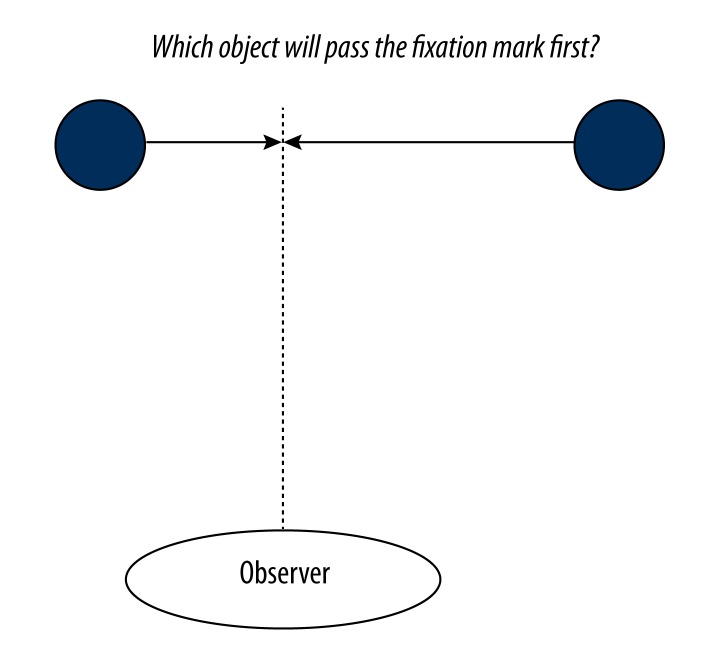
We compared the functional brain connectivity produced during resting-state in which subjects were not actively engaged in a task with that produced while they actively performed a visual motion task (task-state).
In this paper we employed graph-theoretical measures and network statistics in novel ways to compare, in the same group of human subjects, functional brain connectivity during resting-state fMRI with brain connectivity during performance of a high level visual task. We performed a whole-brain connectivity analysis to compare network statistics in resting and task states among anatomically defined Brodmann areas to investigate how brain networks spanning the cortex changed when subjects were engaged in task performance.
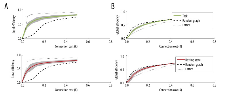
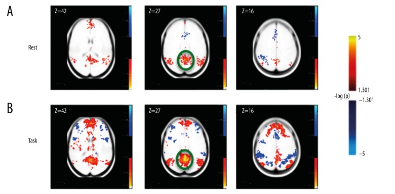
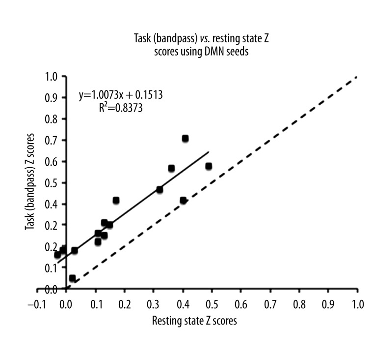
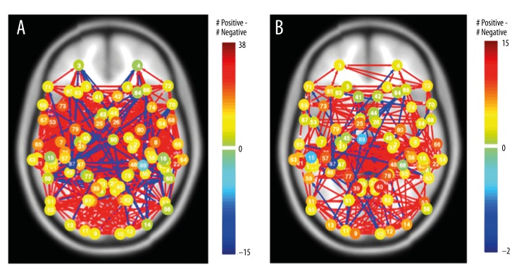
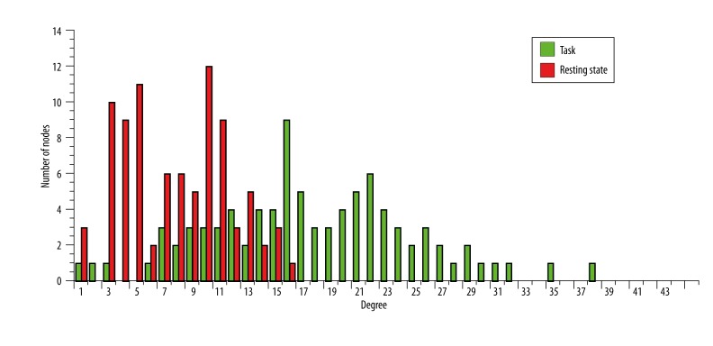
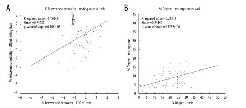
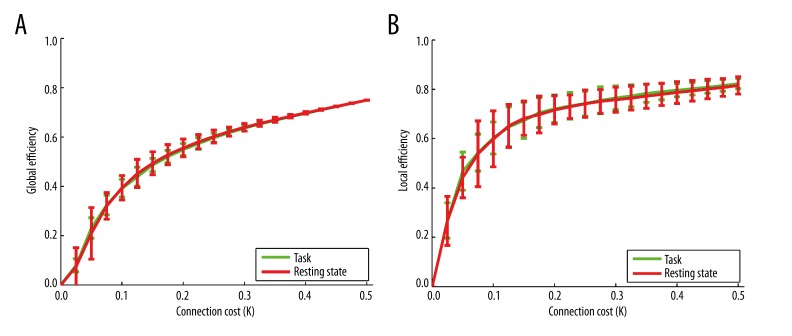
In the resting state, we found strong connectivity among the posterior cingulate cortex (PCC), precuneus, medial prefrontal cortex (MPFC), lateral parietal cortex, and hippocampal formation, consistent with previous reports of the default mode network (DMN). The connections among these areas were strengthened while subjects actively performed an event-related visual motion task, indicating a continued and strong engagement of the DMN during task processing. Regional measures such as degree (number of connections) and betweenness centrality (number of shortest paths), showed that task performance induces stronger inter-regional connections, leading to a denser processing network, but that this does not imply a more efficient system as shown by the integration measures such as path length and global efficiency, and from global measures such as small-worldness.
In spite of the maintenance of connectivity and the "hub-like" behavior of areas, our results suggest that the network paths may be rerouted when performing the task condition.
我们比较了受试者在静息状态(未积极参与任务)下产生的大脑功能连接与他们积极执行视觉运动任务(任务状态)时产生的大脑功能连接。
在本文中,我们以新颖的方式采用图论测量和网络统计方法,在同一组人类受试者中比较静息态功能磁共振成像期间的大脑功能连接与执行高级视觉任务期间的大脑连接。我们进行了全脑连接性分析,以比较解剖学定义的布罗德曼区域在静息和任务状态下的网络统计数据,以研究当受试者参与任务执行时跨越皮质的大脑网络如何变化。
在静息状态下,我们发现后扣带回皮质(PCC)、楔前叶、内侧前额叶皮质(MPFC)、外侧顶叶皮质和海马结构之间存在强连接,这与先前关于默认模式网络(DMN)的报道一致。在受试者积极执行与事件相关的视觉运动任务时,这些区域之间的连接得到加强,表明在任务处理过程中DMN持续且强烈地参与。诸如度(连接数)和介数中心性(最短路径数)等区域测量表明,任务执行会诱导更强的区域间连接,导致处理网络更密集,但如路径长度和全局效率等整合测量以及小世界性质等全局测量所示,这并不意味着系统更高效。
尽管连接性得以维持且区域表现出“中心样”行为,但我们的结果表明,在执行任务时网络路径可能会重新布线。