Bianchini Paolo, Cardarelli Francesco, Di Luca Mariagrazia, Diaspro Alberto, Bizzarri Ranieri
Nanophysics, IIT-Italian Institute of Technology, Genoa, Italy.
Center for Nanotechnology Innovation @NEST, Istituto Italiano di Tecnologia, Pisa, Italy.
PLoS One. 2014 Jun 26;9(6):e99619. doi: 10.1371/journal.pone.0099619. eCollection 2014.
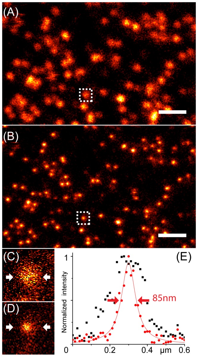
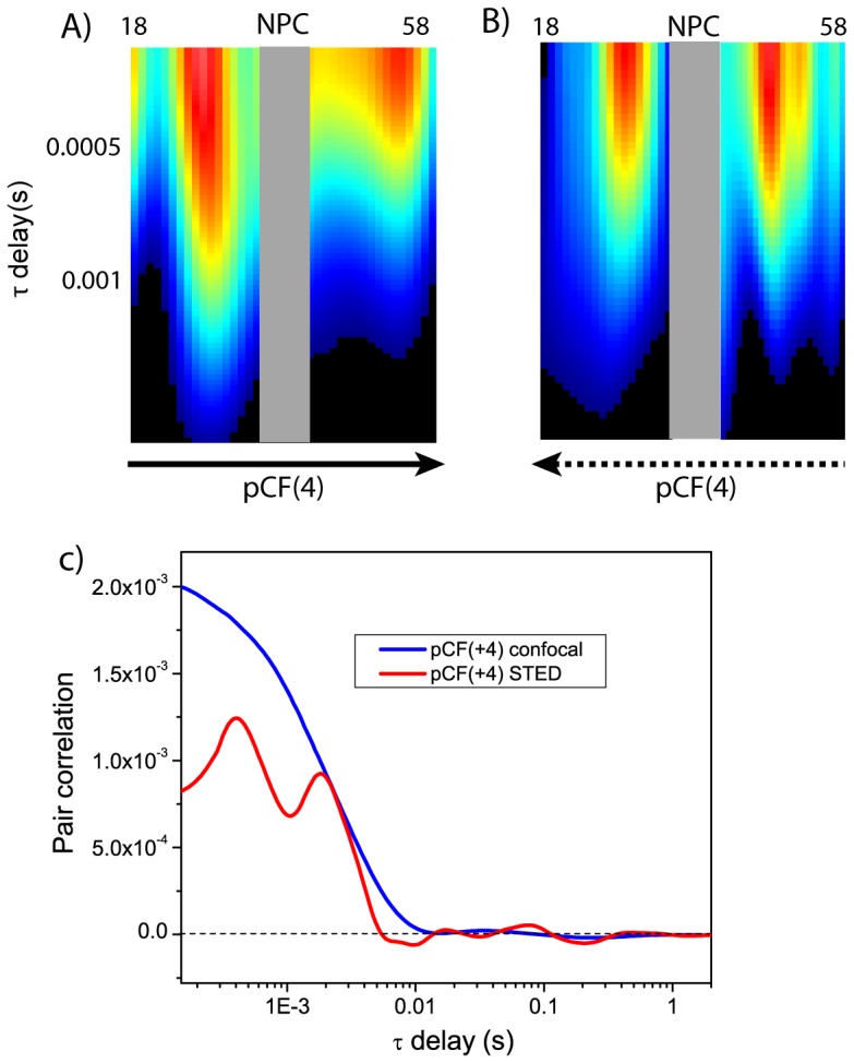
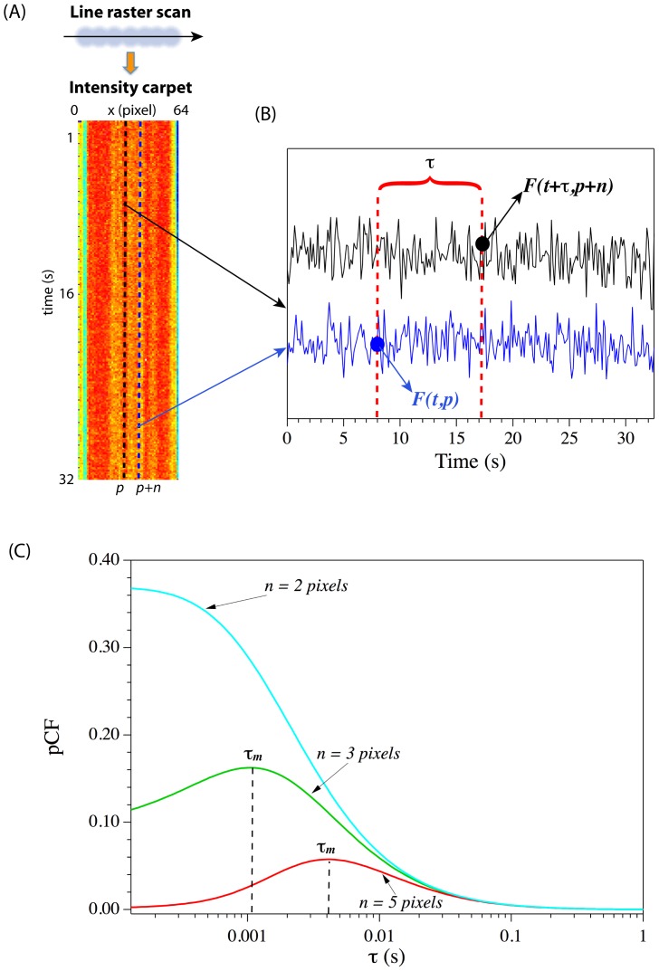
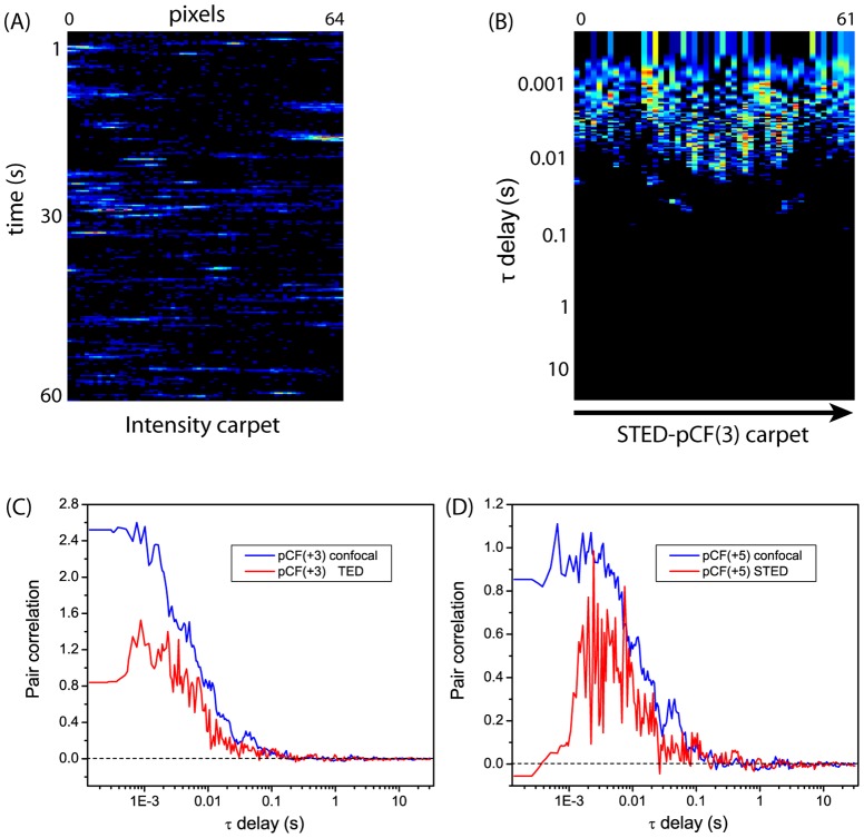
We describe for the first time the combination between cross-pair correlation function analysis (pair correlation analysis or pCF) and stimulated emission depletion (STED) to obtain diffusion maps at spatial resolution below the optical diffraction limit (super-resolution). Our approach was tested in systems characterized by high and low signal to noise ratio, i.e. Capsid Like Particles (CLPs) bearing several (>100) active fluorescent proteins and monomeric fluorescent proteins transiently expressed in living Chinese Hamster Ovary cells, respectively. The latter system represents the usual condition encountered in living cell studies on fluorescent protein chimeras. Spatial resolution of STED-pCF was found to be about 110 nm, with a more than twofold improvement over conventional confocal acquisition. We successfully applied our method to highlight how the proximity to nuclear envelope affects the mobility features of proteins actively imported into the nucleus in living cells. Remarkably, STED-pCF unveiled the existence of local barriers to diffusion as well as the presence of a slow component at distances up to 500-700 nm from either sides of nuclear envelope. The mobility of this component is similar to that previously described for transport complexes. Remarkably, all these features were invisible in conventional confocal mode.
我们首次描述了交叉对相关函数分析(配对相关分析或pCF)与受激发射损耗(STED)相结合的方法,以获得低于光学衍射极限(超分辨率)的空间分辨率下的扩散图。我们的方法在具有高信噪比和低信噪比的系统中进行了测试,即分别在携带几种(>100)活性荧光蛋白的衣壳样颗粒(CLP)和在活的中国仓鼠卵巢细胞中瞬时表达的单体荧光蛋白中进行测试。后一种系统代表了在荧光蛋白嵌合体的活细胞研究中通常遇到的情况。发现STED-pCF的空间分辨率约为110 nm,比传统共聚焦采集提高了两倍多。我们成功地应用我们的方法来突出与核膜的接近程度如何影响活细胞中主动导入细胞核的蛋白质的迁移特征。值得注意的是,STED-pCF揭示了扩散的局部屏障的存在以及在距核膜两侧高达500-700 nm的距离处存在缓慢成分。该成分的迁移率与先前描述的转运复合物的迁移率相似。值得注意的是,所有这些特征在传统共聚焦模式下都是不可见的。