Department of Zoology, College of Science, King Saud University, Saudi Arabia, Riyadh, KSA ; Department of Zoology, Faculty of Science, El-Minia University, El-Minia, Egypt.
Nutr Metab (Lond). 2014 Jul 1;11:31. doi: 10.1186/1743-7075-11-31. eCollection 2014.
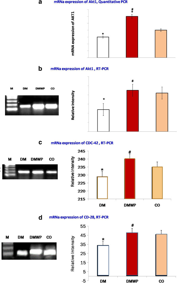
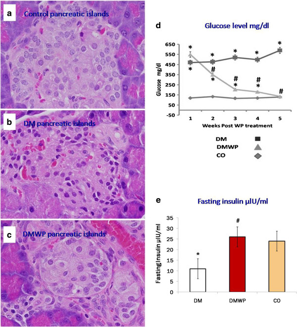
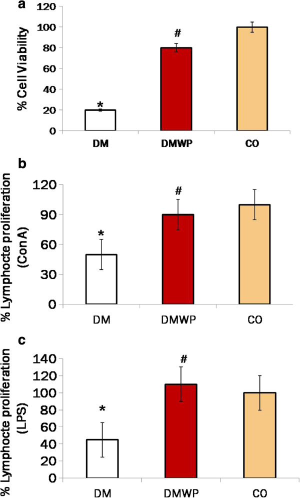
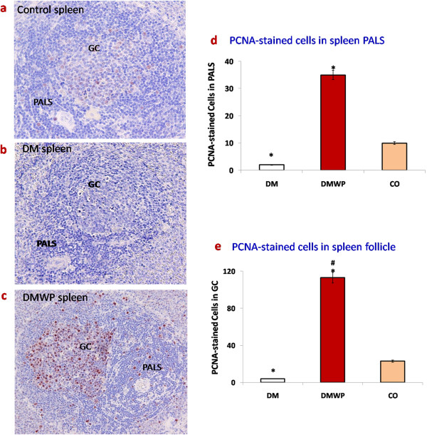
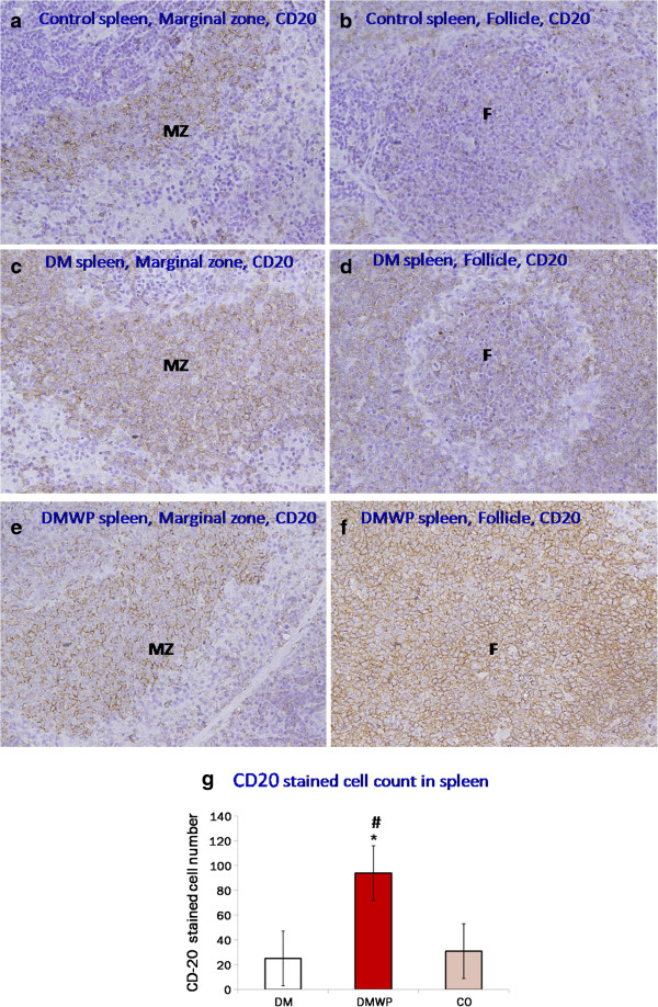
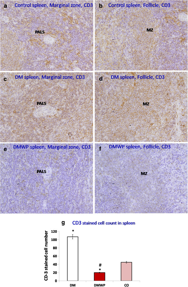
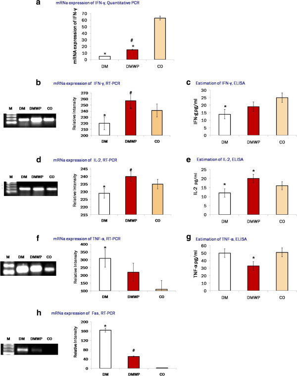
T cell mediated autoimmune diabetes is characterized by immune cell infiltration of pancreatic islets and destruction of insulin-producing β-cells. This study was designed to assess the effect of whey proteins (WP) on the responsiveness of lymphocytes in rats after four months of Streptozotocin (STZ)-induced Type 1 diabetes (T1D). A diabetic group was supplemented with WP daily for five weeks at a dose of 100 mg/kg. Ribonucleic acid (RNA) was extracted from stimulated lymphocytes in order to analyse gene expressions using real time PCR and RT-PCR. PCR results were confirmed with ELISA. The proliferation capacity of lymphocytes and their homing to the spleen were studied. Antigen-activated lymphocytes showed that diabetes impaired the mRNA expression of the protein kinase B (Akt1), Cdc42, and the co-stimulatory molecule, CD28, which are important for cell survival, actin polymerization and T cell activation, respectively. Accordingly, proliferation of lymphocytes was found to be suppressed in diabetic rats, both in vivo and in vitro. WP was found to restore Akt1, Cdc42 and CD28 mRNA expression during diabetes to normal levels. WP, therefore, served to activate the proliferation of B lymphocytes in diabetic rats both in vivo and in vitro. Although WP was found to up-regulate mRNA expression of both interleukin (IL)-2 and interferon gamma (IFN-γ), it suppressed the proliferation activity of almost all T cell subsets. This was confirmed by WP normalizing the structure and function of ß cells. Meanwhile, WP was found to down regulate the mRNA expression of Tumor necrosis factor-alpha (TNF-α) and its programmed cell death-receptor (Fas). Taken together, the results of this study provide evidence for the potential impact of WP in the treatment of immune impairment in T1D, suggesting that it serves to reverse autoimmunity by suppressing autoreactive T cells and down regulating TNF-α and Fas, resulting in improved pancreatic ß cell structure and function.
细胞介导的自身免疫性糖尿病的特征是免疫细胞浸润胰岛和破坏胰岛素分泌β细胞。本研究旨在评估乳清蛋白(WP)对四氯化碳诱导的 1 型糖尿病(T1D)大鼠淋巴细胞反应性的影响。糖尿病组每天补充 WP,剂量为 100mg/kg,持续 5 周。从刺激的淋巴细胞中提取核糖核酸(RNA),以使用实时 PCR 和 RT-PCR 分析基因表达。用 ELISA 验证 PCR 结果。研究了淋巴细胞的增殖能力及其向脾脏的归巢。抗原激活的淋巴细胞表明,糖尿病损害了蛋白激酶 B(Akt1)、Cdc42 和共刺激分子 CD28 的 mRNA 表达,这些对细胞存活、肌动蛋白聚合和 T 细胞激活分别很重要。因此,发现糖尿病大鼠的淋巴细胞增殖受到抑制,无论是在体内还是体外。WP 被发现可在糖尿病期间将 Akt1、Cdc42 和 CD28 的 mRNA 表达恢复到正常水平。因此,WP 可激活糖尿病大鼠体内和体外 B 淋巴细胞的增殖。尽管 WP 被发现可上调白细胞介素(IL)-2 和干扰素γ(IFN-γ)的 mRNA 表达,但它抑制了几乎所有 T 细胞亚群的增殖活性。这一点通过 WP 使 β 细胞的结构和功能正常化得到了证实。同时,WP 被发现可下调肿瘤坏死因子-α(TNF-α)及其程序性细胞死亡受体(Fas)的 mRNA 表达。总之,本研究结果为 WP 在治疗 T1D 免疫损伤中的潜在影响提供了证据,表明 WP 通过抑制自身反应性 T 细胞和下调 TNF-α 和 Fas 来逆转自身免疫,从而改善胰腺 β 细胞的结构和功能。