Shi Jie, Stonnington Cynthia M, Thompson Paul M, Chen Kewei, Gutman Boris, Reschke Cole, Baxter Leslie C, Reiman Eric M, Caselli Richard J, Wang Yalin
School of Computing, Informatics, and Decision Systems Engineering, Arizona State University, Tempe, AZ, USA.
Department of Psychiatry and Psychology, Mayo Clinic Arizona, Scottsdale, AZ, USA.
Neuroimage. 2015 Jan 1;104:1-20. doi: 10.1016/j.neuroimage.2014.09.062. Epub 2014 Oct 5.
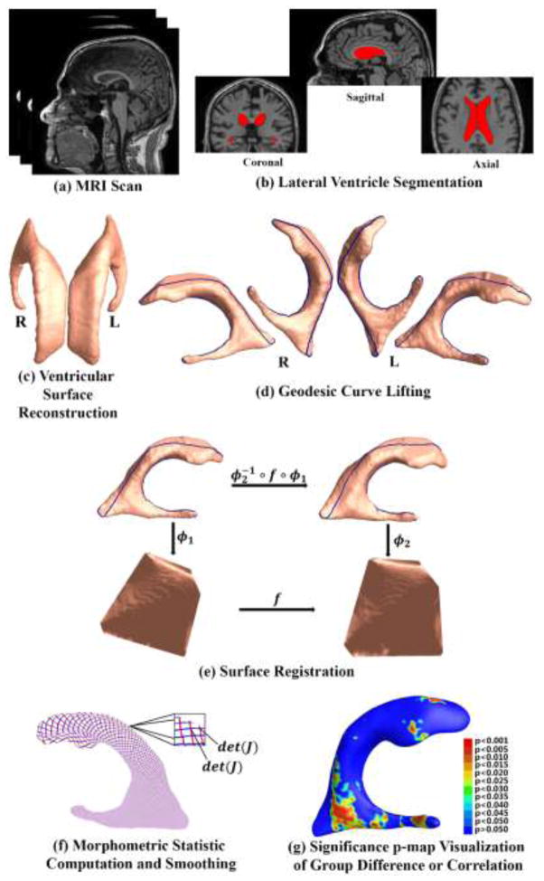
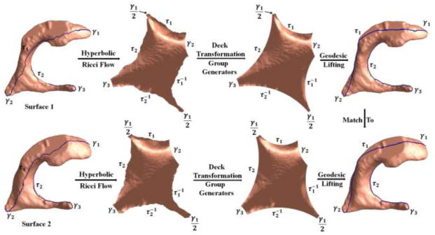
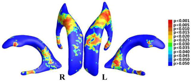
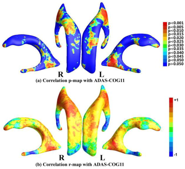
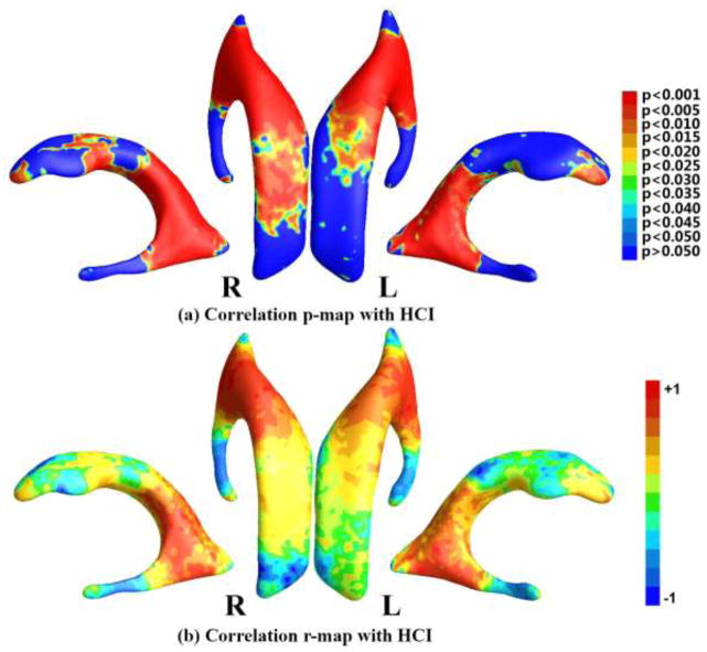
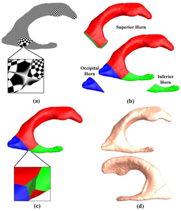
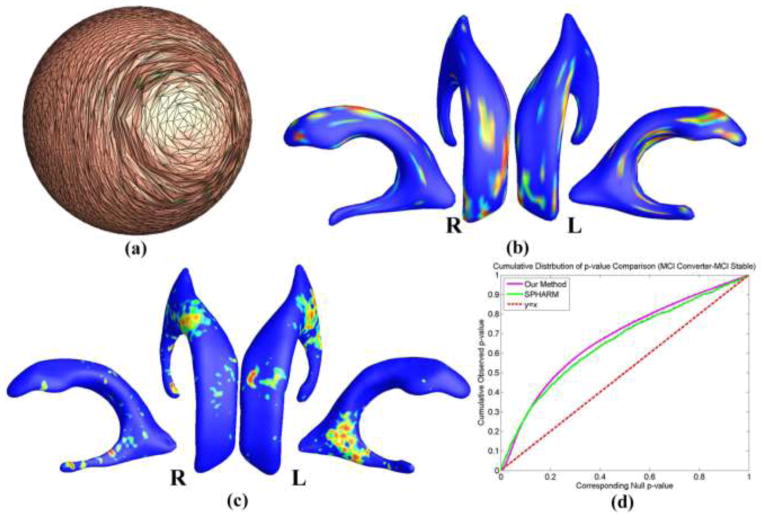
Mild Cognitive Impairment (MCI) is a transitional stage between normal aging and dementia and people with MCI are at high risk of progression to dementia. MCI is attracting increasing attention, as it offers an opportunity to target the disease process during an early symptomatic stage. Structural magnetic resonance imaging (MRI) measures have been the mainstay of Alzheimer's disease (AD) imaging research, however, ventricular morphometry analysis remains challenging because of its complicated topological structure. Here we describe a novel ventricular morphometry system based on the hyperbolic Ricci flow method and tensor-based morphometry (TBM) statistics. Unlike prior ventricular surface parameterization methods, hyperbolic conformal parameterization is angle-preserving and does not have any singularities. Our system generates a one-to-one diffeomorphic mapping between ventricular surfaces with consistent boundary matching conditions. The TBM statistics encode a great deal of surface deformation information that could be inaccessible or overlooked by other methods. We applied our system to the baseline MRI scans of a set of MCI subjects from the Alzheimer's Disease Neuroimaging Initiative (ADNI: 71 MCI converters vs. 62 MCI stable). Although the combined ventricular area and volume features did not differ between the two groups, our fine-grained surface analysis revealed significant differences in the ventricular regions close to the temporal lobe and posterior cingulate, structures that are affected early in AD. Significant correlations were also detected between ventricular morphometry, neuropsychological measures, and a previously described imaging index based on fluorodeoxyglucose positron emission tomography (FDG-PET) scans. This novel ventricular morphometry method may offer a new and more sensitive approach to study preclinical and early symptomatic stage AD.
轻度认知障碍(MCI)是正常衰老与痴呆之间的过渡阶段,患有MCI的人发展为痴呆的风险很高。MCI正吸引着越来越多的关注,因为它为在早期症状阶段针对疾病进程提供了一个机会。结构磁共振成像(MRI)测量一直是阿尔茨海默病(AD)成像研究的主要手段,然而,由于脑室形态测量分析的拓扑结构复杂,其分析仍然具有挑战性。在此,我们描述了一种基于双曲里奇流方法和基于张量的形态测量(TBM)统计的新型脑室形态测量系统。与先前的脑室表面参数化方法不同,双曲共形参数化是保角的,并且没有任何奇点。我们的系统在脑室表面之间生成具有一致边界匹配条件的一一微分同胚映射。TBM统计编码了大量的表面变形信息,而这些信息可能是其他方法无法获取或忽略的。我们将我们的系统应用于来自阿尔茨海默病神经影像倡议(ADNI)的一组MCI受试者的基线MRI扫描(71名MCI转化者与62名MCI稳定者)。尽管两组之间的脑室总面积和体积特征没有差异,但我们的细粒度表面分析显示,靠近颞叶和后扣带回的脑室区域存在显著差异,这些结构在AD早期就会受到影响。在脑室形态测量、神经心理学测量以及先前基于氟脱氧葡萄糖正电子发射断层扫描(FDG-PET)扫描描述的成像指标之间也检测到了显著相关性。这种新型脑室形态测量方法可能为研究临床前和早期症状阶段的AD提供一种新的、更敏感的方法。