Section on Functional Imaging Methods, LBC, NIMH, NIH, Bethesda, MD, USA.
Section on Functional Imaging Methods, LBC, NIMH, NIH, Bethesda, MD, USA.
Neuroimage. 2015 Jan 15;105:189-97. doi: 10.1016/j.neuroimage.2014.10.051. Epub 2014 Oct 30.
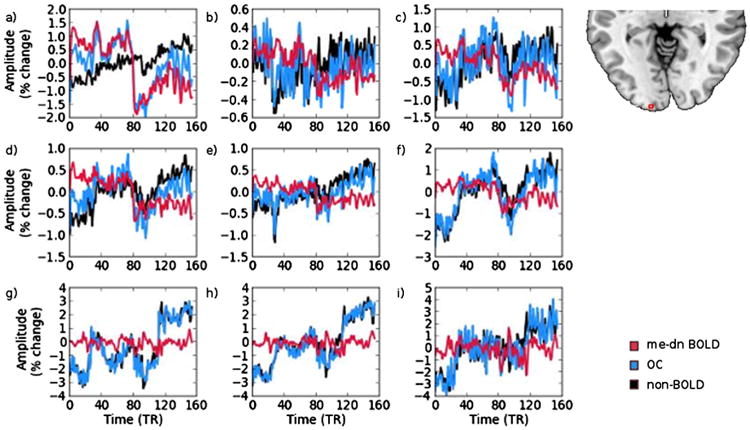
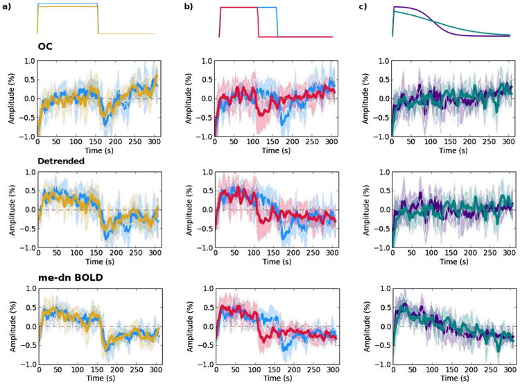
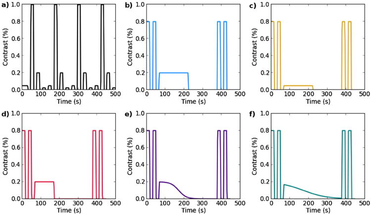
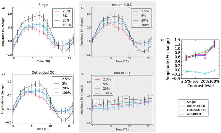
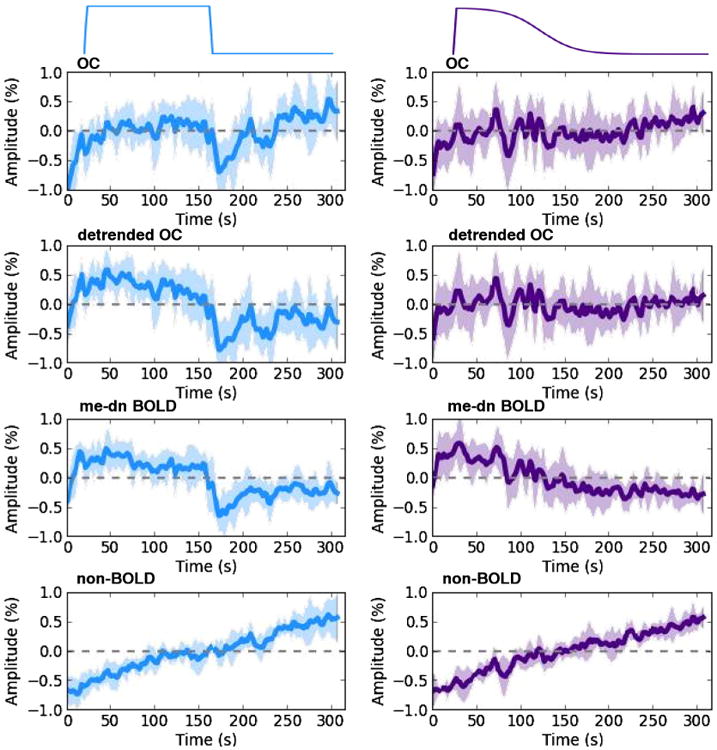
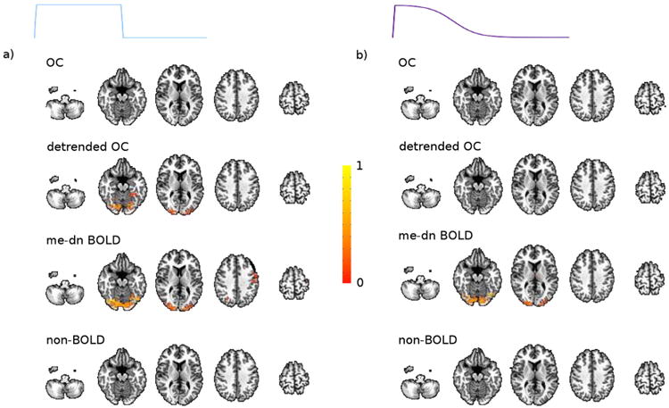
The functional magnetic resonance (fMRI) baseline is known to drift over the course of an experiment and is often attributed to hardware instability. These ultraslow fMRI fluctuations are inseparable from blood oxygenation level dependent (BOLD) changes in standard single echo fMRI and they are therefore typically removed before further analysis in both resting-state and task paradigms. However, some part of these fluctuations may be of neuronal origin, as neural activity can indeed fluctuate at the scale of several minutes or even longer, such as after the administration of drugs or during the ultradian rhythms. Here, we show that it is possible to separate the slow BOLD and non-BOLD drifts automatically using multi-echo fMRI and multi-echo independent components analysis (ME-ICA) denoising by demonstrating the detection of a visual signal evoked from a flickering checkerboard with slowly changing contrast.
功能磁共振成像(fMRI)的基线已知会在实验过程中漂移,通常归因于硬件不稳定。这些超慢 fMRI 波动与标准单回波 fMRI 中的血氧水平依赖(BOLD)变化是不可分割的,因此在静息状态和任务范式中进一步分析之前,通常会将其去除。然而,这些波动的某些部分可能具有神经元起源,因为神经活动实际上可以以数分钟甚至更长的时间尺度波动,例如在药物给药后或在超昼夜节律期间。在这里,我们通过证明可以检测到从闪烁的棋盘上以缓慢变化的对比度诱发的视觉信号,使用多回波 fMRI 和多回波独立成分分析(ME-ICA)去噪自动分离缓慢的 BOLD 和非 BOLD 漂移,表明了这一点。