• 文献检索
  • 文档翻译
  • 深度研究
  • 学术资讯
  • Suppr Zotero 插件Zotero 插件
  • 邀请有礼
  • 套餐&价格
  • 历史记录
应用&插件
Suppr Zotero 插件Zotero 插件浏览器插件Mac 客户端Windows 客户端微信小程序
定价
高级版会员购买积分包购买API积分包
服务
文献检索文档翻译深度研究API 文档MCP 服务
关于我们
关于 Suppr公司介绍联系我们用户协议隐私条款
关注我们

Suppr 超能文献

核心技术专利:CN118964589B侵权必究
粤ICP备2023148730 号-1Suppr @ 2026

文献检索

告别复杂PubMed语法,用中文像聊天一样搜索,搜遍4000万医学文献。AI智能推荐,让科研检索更轻松。

立即免费搜索

文件翻译

保留排版,准确专业,支持PDF/Word/PPT等文件格式,支持 12+语言互译。

免费翻译文档

深度研究

AI帮你快速写综述,25分钟生成高质量综述,智能提取关键信息,辅助科研写作。

立即免费体验

轴突中周期性膜骨架的发育机制。

Developmental mechanism of the periodic membrane skeleton in axons.

作者信息

Zhong Guisheng, He Jiang, Zhou Ruobo, Lorenzo Damaris, Babcock Hazen P, Bennett Vann, Zhuang Xiaowei

机构信息

Department of Chemistry and Chemical Biology, Howard Hughes Medical Institute, Harvard University, Cambridge, United States.

Department of Molecular and Cellular Biology, Howard Hughes Medical Institute, Harvard University, Cambridge, United States.

出版信息

Elife. 2014 Dec 23;3:e04581. doi: 10.7554/eLife.04581.

DOI:10.7554/eLife.04581
PMID:25535840
原文链接:https://pmc.ncbi.nlm.nih.gov/articles/PMC4337613/
Abstract

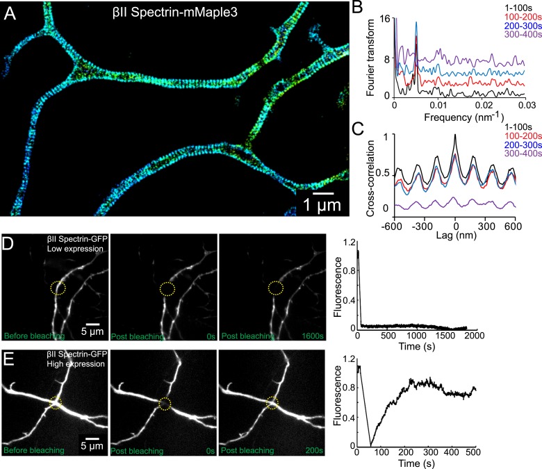
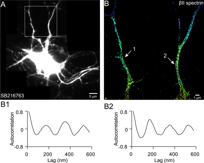
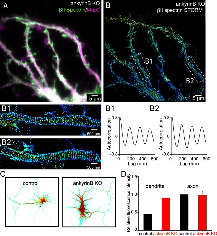
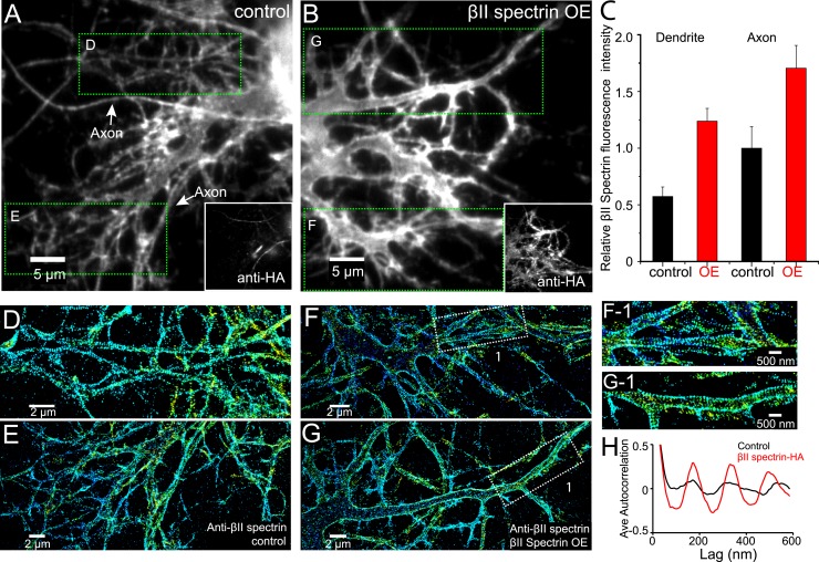
Actin, spectrin, and associated molecules form a periodic sub-membrane lattice structure in axons. How this membrane skeleton is developed and why it preferentially forms in axons are unknown. Here, we studied the developmental mechanism of this lattice structure. We found that this structure emerged early during axon development and propagated from proximal regions to distal ends of axons. Components of the axon initial segment were recruited to the lattice late during development. Formation of the lattice was regulated by the local concentration of βII spectrin, which is higher in axons than in dendrites. Increasing the dendritic concentration of βII spectrin by overexpression or by knocking out ankyrin B induced the formation of the periodic structure in dendrites, demonstrating that the spectrin concentration is a key determinant in the preferential development of this structure in axons and that ankyrin B is critical for the polarized distribution of βII spectrin in neurites.

摘要

肌动蛋白、血影蛋白及相关分子在轴突中形成周期性的亚膜晶格结构。这种膜骨架是如何发育形成的,以及为何它优先在轴突中形成,目前尚不清楚。在此,我们研究了这种晶格结构的发育机制。我们发现,这种结构在轴突发育早期出现,并从轴突的近端区域向远端延伸。轴突起始段的成分在发育后期才被招募到晶格中。晶格的形成受βII血影蛋白局部浓度的调控,轴突中的βII血影蛋白浓度高于树突。通过过表达或敲除锚蛋白B来提高树突中βII血影蛋白的浓度,可诱导树突中形成周期性结构,这表明血影蛋白浓度是该结构在轴突中优先发育的关键决定因素,且锚蛋白B对βII血影蛋白在神经突中的极性分布至关重要。

https://cdn.ncbi.nlm.nih.gov/pmc/blobs/8bb1/4337613/2930fba898cb/elife04581f010.jpg
https://cdn.ncbi.nlm.nih.gov/pmc/blobs/8bb1/4337613/292f08fdc4bd/elife04581f001.jpg
https://cdn.ncbi.nlm.nih.gov/pmc/blobs/8bb1/4337613/bf35b00d26da/elife04581fs001.jpg
https://cdn.ncbi.nlm.nih.gov/pmc/blobs/8bb1/4337613/7a02364bae94/elife04581fs002.jpg
https://cdn.ncbi.nlm.nih.gov/pmc/blobs/8bb1/4337613/0faa2e40c286/elife04581fs003.jpg
https://cdn.ncbi.nlm.nih.gov/pmc/blobs/8bb1/4337613/d2710f56842f/elife04581fs004.jpg
https://cdn.ncbi.nlm.nih.gov/pmc/blobs/8bb1/4337613/c2e52bc3f2ed/elife04581fs005.jpg
https://cdn.ncbi.nlm.nih.gov/pmc/blobs/8bb1/4337613/48413ba6c8a4/elife04581fs006.jpg
https://cdn.ncbi.nlm.nih.gov/pmc/blobs/8bb1/4337613/c4c25c62a3ab/elife04581f002.jpg
https://cdn.ncbi.nlm.nih.gov/pmc/blobs/8bb1/4337613/5486a96e6cb9/elife04581f003.jpg
https://cdn.ncbi.nlm.nih.gov/pmc/blobs/8bb1/4337613/780059bc639e/elife04581fs007.jpg
https://cdn.ncbi.nlm.nih.gov/pmc/blobs/8bb1/4337613/5f82185b9106/elife04581f004.jpg
https://cdn.ncbi.nlm.nih.gov/pmc/blobs/8bb1/4337613/f60a2cf84e96/elife04581fs008.jpg
https://cdn.ncbi.nlm.nih.gov/pmc/blobs/8bb1/4337613/02f5a453b419/elife04581fs009.jpg
https://cdn.ncbi.nlm.nih.gov/pmc/blobs/8bb1/4337613/a212d3416d4f/elife04581f005.jpg
https://cdn.ncbi.nlm.nih.gov/pmc/blobs/8bb1/4337613/ff0e226bec51/elife04581fs010.jpg
https://cdn.ncbi.nlm.nih.gov/pmc/blobs/8bb1/4337613/8751c7311b99/elife04581f006.jpg
https://cdn.ncbi.nlm.nih.gov/pmc/blobs/8bb1/4337613/221d7b5cafc2/elife04581fs011.jpg
https://cdn.ncbi.nlm.nih.gov/pmc/blobs/8bb1/4337613/649217a60fda/elife04581f007.jpg
https://cdn.ncbi.nlm.nih.gov/pmc/blobs/8bb1/4337613/302f2beed59c/elife04581fs012.jpg
https://cdn.ncbi.nlm.nih.gov/pmc/blobs/8bb1/4337613/eb29320d5c6b/elife04581fs013.jpg
https://cdn.ncbi.nlm.nih.gov/pmc/blobs/8bb1/4337613/303bfe9c407f/elife04581f008.jpg
https://cdn.ncbi.nlm.nih.gov/pmc/blobs/8bb1/4337613/8475c286797c/elife04581f009.jpg
https://cdn.ncbi.nlm.nih.gov/pmc/blobs/8bb1/4337613/2930fba898cb/elife04581f010.jpg

相似文献

1
Developmental mechanism of the periodic membrane skeleton in axons.轴突中周期性膜骨架的发育机制。
Elife. 2014 Dec 23;3:e04581. doi: 10.7554/eLife.04581.
2
Structural organization of the actin-spectrin-based membrane skeleton in dendrites and soma of neurons.神经元树突和胞体中肌动蛋白-血影蛋白为基础的膜骨架的结构组织。
Proc Natl Acad Sci U S A. 2017 Aug 8;114(32):E6678-E6685. doi: 10.1073/pnas.1705043114. Epub 2017 Jul 24.
3
Prevalent presence of periodic actin-spectrin-based membrane skeleton in a broad range of neuronal cell types and animal species.周期性肌动蛋白-血影蛋白膜骨架在广泛的神经元细胞类型和动物物种中普遍存在。
Proc Natl Acad Sci U S A. 2016 May 24;113(21):6029-34. doi: 10.1073/pnas.1605707113. Epub 2016 May 9.
4
Spectrin- and ankyrin-based membrane domains and the evolution of vertebrates.基于 spectrin 和 ankyrin 的膜结构域与脊椎动物的演化。
Curr Top Membr. 2013;72:1-37. doi: 10.1016/B978-0-12-417027-8.00001-5.
5
An Adaptable Spectrin/Ankyrin-Based Mechanism for Long-Range Organization of Plasma Membranes in Vertebrate Tissues.一种基于血影蛋白/锚蛋白的适应性机制,用于脊椎动物组织中质膜的长距离组织。
Curr Top Membr. 2016;77:143-84. doi: 10.1016/bs.ctm.2015.10.001. Epub 2015 Nov 30.
6
βII-spectrin promotes mouse brain connectivity through stabilizing axonal plasma membranes and enabling axonal organelle transport.βII- spectrin 通过稳定轴突质膜和促进轴突细胞器运输来促进小鼠大脑连接。
Proc Natl Acad Sci U S A. 2019 Jul 30;116(31):15686-15695. doi: 10.1073/pnas.1820649116. Epub 2019 Jun 17.
7
Structural plasticity of actin-spectrin membrane skeleton and functional role of actin and spectrin in axon degeneration.肌动蛋白-血影蛋白膜骨架的结构可塑性及肌动蛋白和血影蛋白在轴突变性中的功能作用。
Elife. 2019 May 1;8:e38730. doi: 10.7554/eLife.38730.
8
Expression and assembly of the erythroid membrane-skeletal proteins ankyrin (goblin) and spectrin in the morphogenesis of chicken neurons.红细胞膜骨架蛋白锚蛋白(地精蛋白)和血影蛋白在鸡神经元形态发生中的表达与组装。
J Cell Biochem. 1985;27(4):423-41. doi: 10.1002/jcb.240270411.
9
Modeling of the axon membrane skeleton structure and implications for its mechanical properties.轴突膜骨架结构建模及其力学性能研究
PLoS Comput Biol. 2017 Feb 27;13(2):e1005407. doi: 10.1371/journal.pcbi.1005407. eCollection 2017 Feb.
10
The role of the membrane-associated periodic skeleton in axons.膜相关周期骨架在轴突中的作用。
Cell Mol Life Sci. 2021 Jul;78(13):5371-5379. doi: 10.1007/s00018-021-03867-x. Epub 2021 Jun 3.

引用本文的文献

1
A correlation-based tool for quantifying membrane periodic skeleton associated periodicity.一种基于相关性的工具,用于量化膜相关周期性骨架的周期性。
Front Neuroinform. 2025 Aug 22;19:1628538. doi: 10.3389/fninf.2025.1628538. eCollection 2025.
2
The membrane skeleton is constitutively remodeled in neurons by calcium signaling.在神经元中,膜骨架通过钙信号传导持续重塑。
Science. 2025 Aug 7;389(6760):eadn6712. doi: 10.1126/science.adn6712.
3
Super-resolution imaging of the neuronal cytoskeleton.神经元细胞骨架的超分辨率成像。

本文引用的文献

1
A PIK3C3-ankyrin-B-dynactin pathway promotes axonal growth and multiorganelle transport.一条PIK3C3-锚蛋白B-动力蛋白激活蛋白途径促进轴突生长和多细胞器运输。
J Cell Biol. 2014 Dec 22;207(6):735-52. doi: 10.1083/jcb.201407063.
2
The contribution of de novo coding mutations to autism spectrum disorder.新生编码突变对自闭症谱系障碍的影响。
Nature. 2014 Nov 13;515(7526):216-21. doi: 10.1038/nature13908. Epub 2014 Oct 29.
3
Synaptic, transcriptional and chromatin genes disrupted in autism.在自闭症中受到破坏的突触、转录和染色质基因。
Npj Imaging. 2024 Dec 4;2(1):50. doi: 10.1038/s44303-024-00054-y.
4
Stressed Actin Binding by the Prickle2 LIM Domains and its Regulation in the Full-Length Protein.由Prickle2 LIM结构域介导的应激肌动蛋白结合及其在全长蛋白中的调控
bioRxiv. 2025 Jun 2:2025.05.30.657073. doi: 10.1101/2025.05.30.657073.
5
Molecular consequences of SCA5 mutations in the spectrin-repeat domains of β-III-spectrin.β-III-血影蛋白的血影蛋白重复结构域中SCA5突变的分子后果。
J Biol Chem. 2025 Jun 6;301(7):110350. doi: 10.1016/j.jbc.2025.110350.
6
Paralemmin-1 controls the nanoarchitecture of the neuronal submembrane cytoskeleton.Paralemmin-1蛋白控制着神经元亚膜细胞骨架的纳米结构。
Sci Adv. 2025 Mar 7;11(10):eadt3724. doi: 10.1126/sciadv.adt3724.
7
Membrane mechanics dictate axonal pearls-on-a-string morphology and function.膜力学决定轴突上珍珠串样形态和功能。
Nat Neurosci. 2025 Jan;28(1):49-61. doi: 10.1038/s41593-024-01813-1. Epub 2024 Dec 2.
8
Axonal mitochondria regulate gentle touch response through control of axonal actin dynamics.轴突线粒体通过控制轴突肌动蛋白动力学来调节轻触觉反应。
bioRxiv. 2024 Aug 13:2024.08.13.607780. doi: 10.1101/2024.08.13.607780.
9
Using ALS to understand profilin 1's diverse roles in cellular physiology.利用肌萎缩侧索硬化症来了解原肌球蛋白结合蛋白1在细胞生理学中的多种作用。
Cytoskeleton (Hoboken). 2025 Mar;82(3):111-129. doi: 10.1002/cm.21896. Epub 2024 Jul 26.
10
Mechanical characterization of spectrin at the molecular level.在分子水平上对血影蛋白进行力学特性分析。
Sci Rep. 2024 Jul 18;14(1):16631. doi: 10.1038/s41598-024-67500-0.
Nature. 2014 Nov 13;515(7526):209-15. doi: 10.1038/nature13772. Epub 2014 Oct 29.
4
Characterization and development of photoactivatable fluorescent proteins for single-molecule-based superresolution imaging.用于单分子超分辨率成像的光激活荧光蛋白的特性和开发。
Proc Natl Acad Sci U S A. 2014 Jun 10;111(23):8452-7. doi: 10.1073/pnas.1406593111. Epub 2014 May 27.
5
Fluorogenic probes for live-cell imaging of the cytoskeleton.细胞骨架的活细胞成像用荧光探针。
Nat Methods. 2014 Jul;11(7):731-3. doi: 10.1038/nmeth.2972. Epub 2014 May 25.
6
Axon initial segment cytoskeleton comprises a multiprotein submembranous coat containing sparse actin filaments.轴突起始段细胞骨架由包含稀疏肌动蛋白丝的多蛋白亚膜包被组成。
J Cell Biol. 2014 Apr 14;205(1):67-81. doi: 10.1083/jcb.201401045. Epub 2014 Apr 7.
7
Mechanical control of the sense of touch by β-spectrin.β- spectrin 对触觉的机械控制。
Nat Cell Biol. 2014 Mar;16(3):224-33. doi: 10.1038/ncb2915. Epub 2014 Feb 23.
8
Spectrin- and ankyrin-based membrane domains and the evolution of vertebrates.基于 spectrin 和 ankyrin 的膜结构域与脊椎动物的演化。
Curr Top Membr. 2013;72:1-37. doi: 10.1016/B978-0-12-417027-8.00001-5.
9
Dual function of CD81 in influenza virus uncoating and budding.CD81 在流感病毒脱壳和出芽中的双重功能。
PLoS Pathog. 2013;9(10):e1003701. doi: 10.1371/journal.ppat.1003701. Epub 2013 Oct 10.
10
Actin, spectrin, and associated proteins form a periodic cytoskeletal structure in axons.肌动蛋白、血影蛋白和相关蛋白在轴突中形成周期性细胞骨架结构。
Science. 2013 Jan 25;339(6118):452-6. doi: 10.1126/science.1232251. Epub 2012 Dec 13.