Tam Johnny, Cordier Guillaume Alan, Bálint Štefan, Sandoval Álvarez Ángel, Borbely Joseph Steven, Lakadamyali Melike
ICFO-Institut de Ciències Fotòniques (ICFO), 08860, Castelledefels (Barcelona), Spain.
PLoS One. 2014 Dec 29;9(12):e115512. doi: 10.1371/journal.pone.0115512. eCollection 2014.
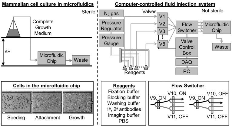
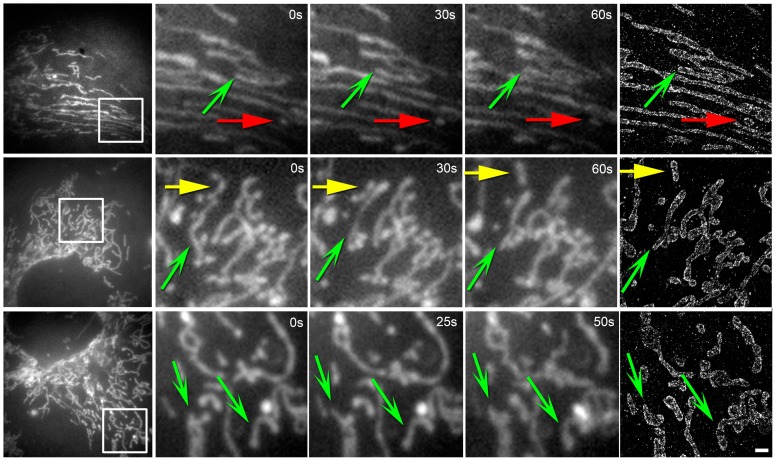
Recently, super-resolution microscopy methods such as stochastic optical reconstruction microscopy (STORM) have enabled visualization of subcellular structures below the optical resolution limit. Due to the poor temporal resolution, however, these methods have mostly been used to image fixed cells or dynamic processes that evolve on slow time-scales. In particular, fast dynamic processes and their relationship to the underlying ultrastructure or nanoscale protein organization cannot be discerned. To overcome this limitation, we have recently developed a correlative and sequential imaging method that combines live-cell and super-resolution microscopy. This approach adds dynamic background to ultrastructural images providing a new dimension to the interpretation of super-resolution data. However, currently, it suffers from the need to carry out tedious steps of sample preparation manually. To alleviate this problem, we implemented a simple and versatile microfluidic platform that streamlines the sample preparation steps in between live-cell and super-resolution imaging. The platform is based on a microfluidic chip with parallel, miniaturized imaging chambers and an automated fluid-injection device, which delivers a precise amount of a specified reagent to the selected imaging chamber at a specific time within the experiment. We demonstrate that this system can be used for live-cell imaging, automated fixation, and immunostaining of adherent mammalian cells in situ followed by STORM imaging. We further demonstrate an application by correlating mitochondrial dynamics, morphology, and nanoscale mitochondrial protein distribution in live and super-resolution images.
最近,诸如随机光学重建显微镜(STORM)等超分辨率显微镜方法能够可视化低于光学分辨率极限的亚细胞结构。然而,由于时间分辨率较差,这些方法大多用于对固定细胞或在缓慢时间尺度上演变的动态过程进行成像。特别是,快速动态过程及其与潜在超微结构或纳米级蛋白质组织的关系无法辨别。为了克服这一限制,我们最近开发了一种将活细胞显微镜和超分辨率显微镜相结合的相关和顺序成像方法。这种方法为超微结构图像添加了动态背景,为超分辨率数据的解释提供了一个新的维度。然而,目前它需要手动执行繁琐的样品制备步骤。为了缓解这个问题,我们实现了一个简单且通用的微流控平台,该平台简化了活细胞和超分辨率成像之间的样品制备步骤。该平台基于一个带有平行、小型化成像室的微流控芯片和一个自动流体注射装置,该装置在实验中的特定时间将精确量的指定试剂输送到选定的成像室。我们证明该系统可用于贴壁哺乳动物细胞的活细胞成像、自动固定和原位免疫染色,随后进行STORM成像。我们进一步通过关联活细胞和超分辨率图像中的线粒体动力学、形态和纳米级线粒体蛋白质分布来展示一种应用。