Behnam Azad Babak, Banerjee Sangeeta R, Pullambhatla Mrudula, Lacerda Silvia, Foss Catherine A, Wang Yuchuan, Ivkov Robert, Pomper Martin G
Russell H. Morgan Department of Radiology and Radiological Science, Johns Hopkins Medical Institutions, Baltimore, MD, USA.
Nanoscale. 2015 Mar 14;7(10):4432-42. doi: 10.1039/c4nr06069e.
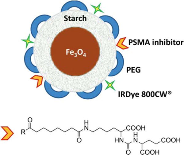
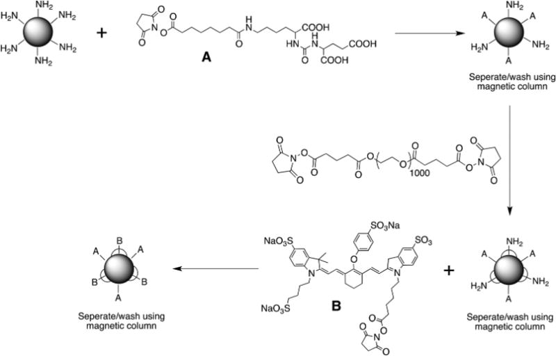
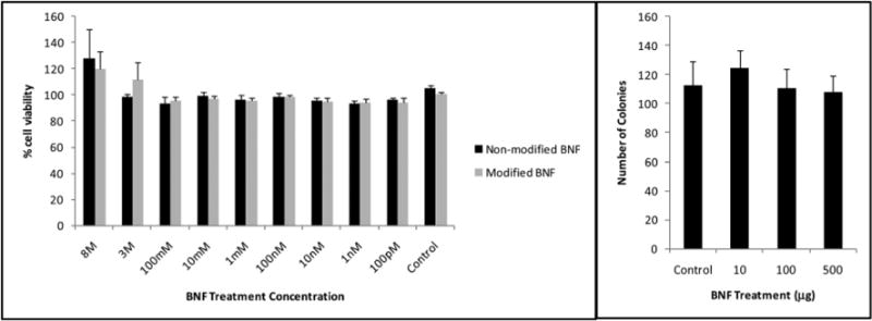
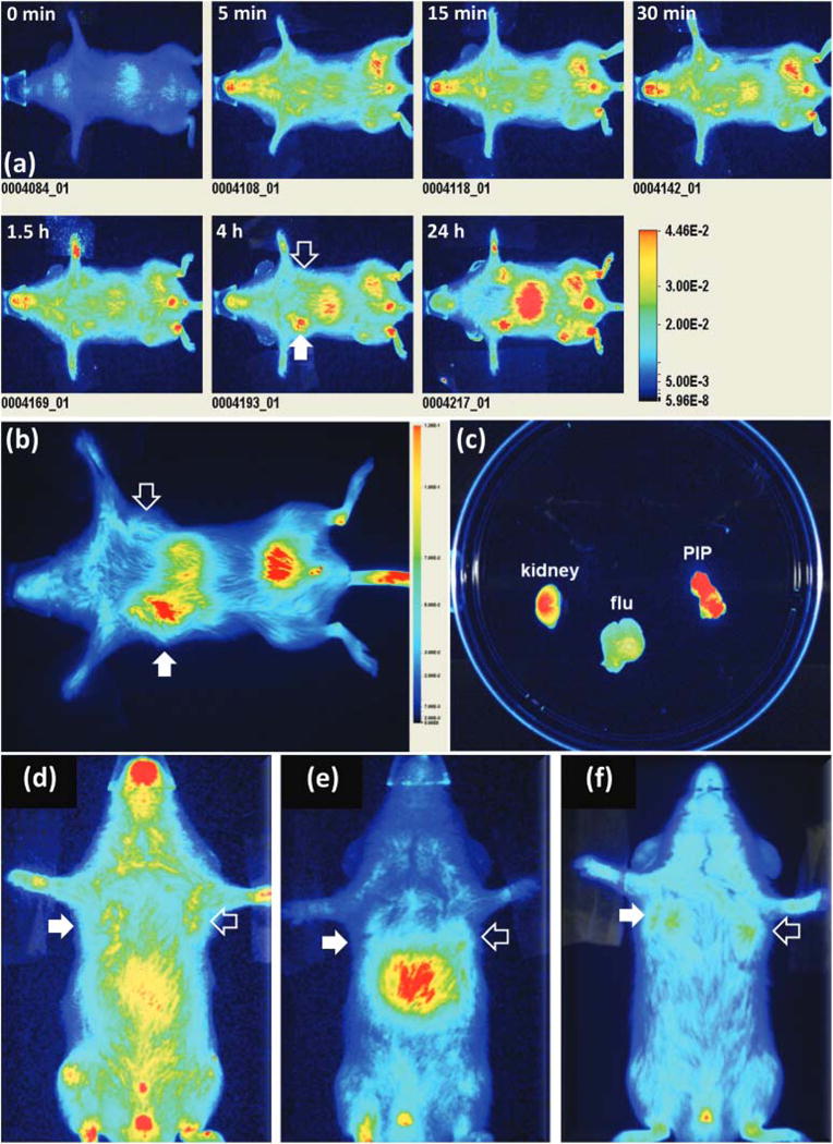
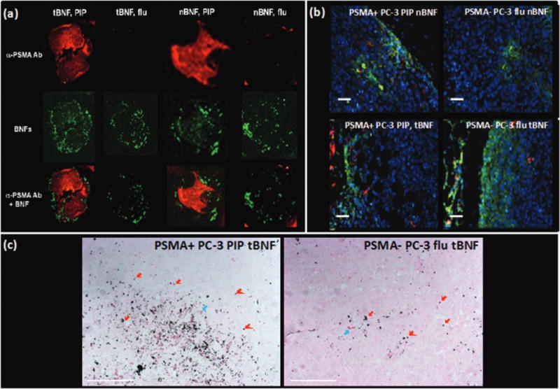
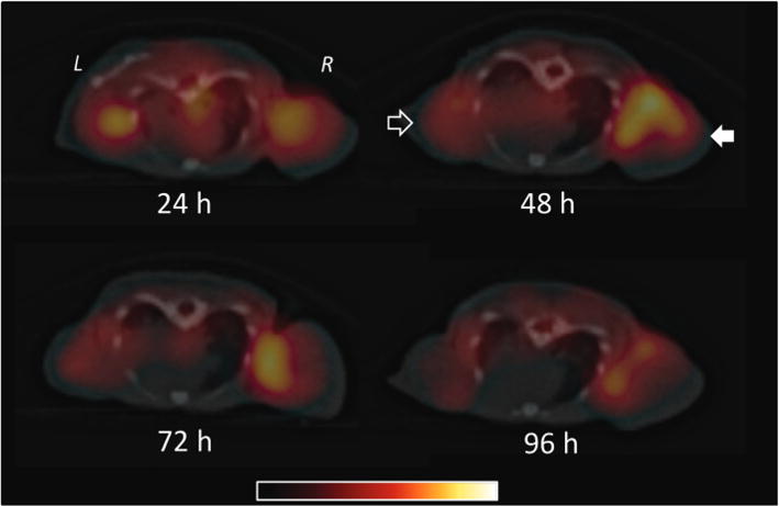
Early detection enables improved prognosis for prostate cancer (PCa). A promising target for imaging and therapy of PCa is the prostate-specific membrane antigen (PSMA), which exhibits both expression within the epithelium of PCa cells, and becomes internalized upon ligand binding. Here we report the synthesis of a PSMA-targeted bionized nanoferrite (BNF) nanoparticle and its biological evaluation in an experimental model of PCa. The BNF nanoparticle formulation exhibits properties conducive to targeted imaging such as stealth, prolonged circulation time and enhanced clearance from non-target sites. Optical imaging of the targeted BNF in vivo indicates preferential accumulation in PSMA+ tumors 4 h post-injection, suggesting target specificity. On the other hand, non-targeted nanoparticles exhibit lower uptake with similar accumulation in both PSMA+ and PSMA- tumors indicating tumor access without preferential accumulation. Imaging with single photon emission computed tomography (SPECT) and biodistribution studies of a modified construct indicate highest tumor accumulation at 48 h post-injection [4.3 ± 0.4 percentage injected dose per gram of tissue (%ID g(-1))], with tumor/blood and tumor/muscle ratios of 7.5 ± 2.4 and 11.6 ± 1.2 %ID g(-1), respectively. Ex vivo fluorescence microscopy, Prussian blue staining, immunohistochemistry and biodistribution studies confirm enhanced nanoparticle uptake in PSMA+ tumors compared to those not expressing PSMA. The BNF nano-formulation described is promising for PSMA-targeted imaging applications in vivo.
早期检测有助于改善前列腺癌(PCa)的预后。前列腺特异性膜抗原(PSMA)是PCa成像和治疗的一个有前景的靶点,它在PCa细胞上皮内表达,并且在配体结合后会内化。在此,我们报告了一种靶向PSMA的生物离子化纳米铁氧体(BNF)纳米颗粒的合成及其在PCa实验模型中的生物学评估。BNF纳米颗粒制剂具有有利于靶向成像的特性,如隐身性、延长的循环时间和从非靶位点的增强清除。靶向BNF在体内的光学成像表明,注射后4小时在PSMA+肿瘤中优先积累,表明具有靶标特异性。另一方面,非靶向纳米颗粒在PSMA+和PSMA-肿瘤中的摄取较低且积累相似,表明可进入肿瘤但无优先积累。单光子发射计算机断层扫描(SPECT)成像和一种修饰构建体的生物分布研究表明,注射后48小时肿瘤积累最高[每克组织注射剂量的4.3±0.4百分比(%ID g(-1))],肿瘤/血液和肿瘤/肌肉比率分别为7.5±2.4和11.6±1.2 %ID g(-1)。体外荧光显微镜、普鲁士蓝染色、免疫组织化学和生物分布研究证实,与未表达PSMA的肿瘤相比,PSMA+肿瘤中纳米颗粒摄取增强。所描述的BNF纳米制剂在体内PSMA靶向成像应用方面很有前景。