Obstoy Bérengère, Salaun Mathieu, Veresezan Liana, Sesboüé Richard, Bohn Pierre, Boland François-Xavier, Thiberville Luc
Quant.I.F Litis EA 4108, IRIB, Rouen University, Rouen, F-76000, France.
Clinique Pneumologique and CIC INSERM U1404, IRIB, Rouen University Hospital, Rouen, F-76031, France.
BMC Pulm Med. 2015 Mar 31;15:30. doi: 10.1186/s12890-015-0020-4.
Fibered confocal fluorescence microscopy (FCFM) allows in vivo investigation of pulmonary microstructures. However, the bronchial epithelium can only be imaged using exogenous fluorophores. The objective of this study is to compare methylene blue (MB) and acriflavine genotoxicity and to assess FCFM performance for in vivo imaging of precancerous lesions.
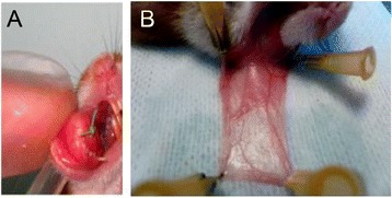
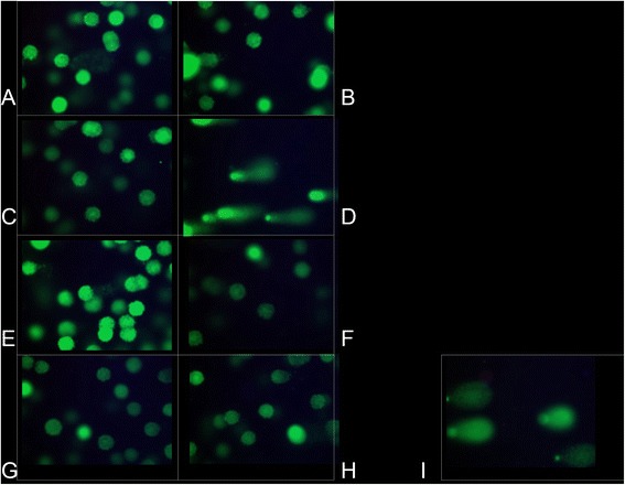
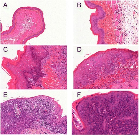
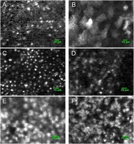
Genotoxicity was assessed using the comet assay on both cultured human lymphocytes and NCI-H460 cells, which had been exposed to MB or acriflavine before being illuminated at 660 or 488 nm, respectively. FCFM was performed on precancerous lesions in the hamster cheek pouch model, following topical application of the fluorophores. FCFM data were analyzed according to histology.
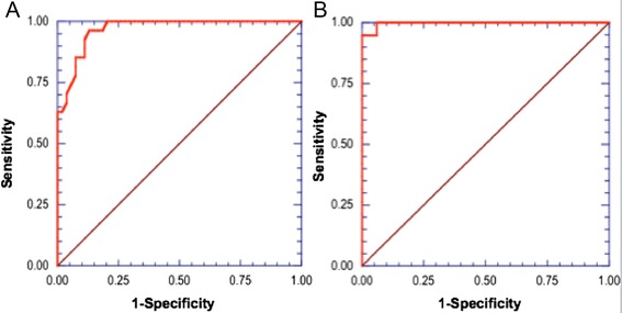
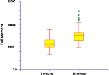
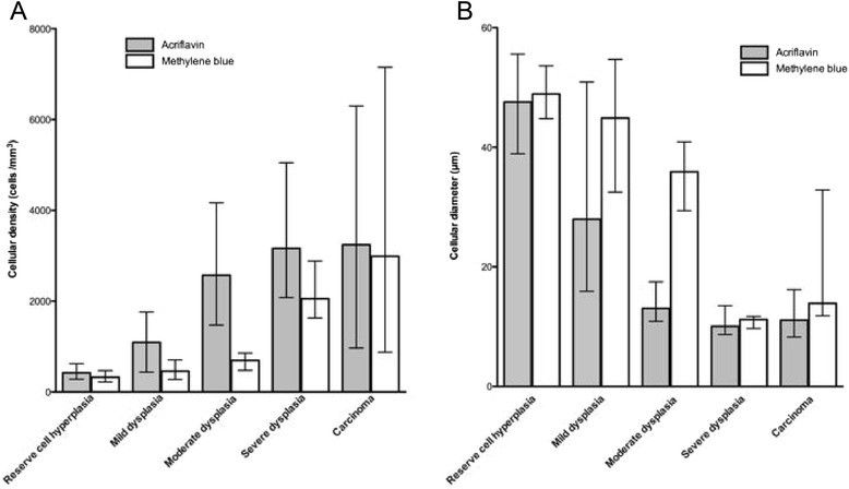
No genotoxicity was found using 0.01% (w/v) MB after illumination at 660 nm for 2 and 15 min (5 mW). Acriflavine exposure (0.025%) led to DNA damages, increasing from 2 to 15 min of light exposure at 448 nm in both lymphocytes (83.4 to 88%, p = 0.021) and NCI H460 cell populations (79.9 to 84.6%, p = 0.045). In total, 11 invasive carcinoma, 24 reserve cell hyperplasia, and 17 dysplasia lesions were imaged using FCFM in vivo. With both fluorophores, the cellular density increased from hyperplasia to high-grade dysplasia (p < 0.05). With MB, the cellular diameter significantly decreased (48.9 to 13.9 μm) from hyperplasia to carcinoma (p < 0.05). In this model, a cut-off diameter of 30 μm enabled the diagnosis of high-grade lesions with a sensitivity of 94.7% and a specificity of 97%.
Methylene blue can be used safely to image precancerous lesions in vivo. This study does not support the use of acriflavine in humans.
纤维共聚焦荧光显微镜(FCFM)可对肺部微观结构进行体内研究。然而,支气管上皮只能使用外源性荧光团成像。本研究的目的是比较亚甲蓝(MB)和吖啶黄的遗传毒性,并评估FCFM对癌前病变进行体内成像的性能。
使用彗星试验评估培养的人淋巴细胞和NCI-H460细胞的遗传毒性,这些细胞在分别于660或488nm光照前暴露于MB或吖啶黄。在仓鼠颊囊模型的癌前病变上进行FCFM,荧光团局部应用后进行。根据组织学分析FCFM数据。
在660nm光照2分钟和15分钟(5mW)后,使用0.01%(w/v)MB未发现遗传毒性。吖啶黄暴露(0.025%)导致DNA损伤,在淋巴细胞(从83.4%增加到88%,p=0.021)和NCI H460细胞群体(从79.9%增加到84.6%,p=0.045)中,448nm光照2分钟到15分钟期间DNA损伤增加。总共使用FCFM对11例浸润性癌、24例储备细胞增生和17例发育异常病变进行了体内成像。使用两种荧光团时,细胞密度从增生到高级别发育异常增加(p<0.05)。使用MB时,细胞直径从增生到癌显著减小(从48.9μm到13.9μm)(p<0.05)。在该模型中,30μm的临界直径能够诊断高级别病变,灵敏度为94.7%,特异性为97%。
亚甲蓝可安全用于体内癌前病变成像。本研究不支持在人体中使用吖啶黄。