• 文献检索
  • 文档翻译
  • 深度研究
  • 学术资讯
  • Suppr Zotero 插件Zotero 插件
  • 邀请有礼
  • 套餐&价格
  • 历史记录
应用&插件
Suppr Zotero 插件Zotero 插件浏览器插件Mac 客户端Windows 客户端微信小程序
定价
高级版会员购买积分包购买API积分包
服务
文献检索文档翻译深度研究API 文档MCP 服务
关于我们
关于 Suppr公司介绍联系我们用户协议隐私条款
关注我们

Suppr 超能文献

核心技术专利:CN118964589B侵权必究
粤ICP备2023148730 号-1Suppr @ 2026

文献检索

告别复杂PubMed语法,用中文像聊天一样搜索,搜遍4000万医学文献。AI智能推荐,让科研检索更轻松。

立即免费搜索

文件翻译

保留排版,准确专业,支持PDF/Word/PPT等文件格式,支持 12+语言互译。

免费翻译文档

深度研究

AI帮你快速写综述,25分钟生成高质量综述,智能提取关键信息,辅助科研写作。

立即免费体验

基于活体共聚焦显微镜的角膜内皮细胞全自动评估

Fully automatic evaluation of the corneal endothelium from in vivo confocal microscopy.

作者信息

Selig Bettina, Vermeer Koenraad A, Rieger Bernd, Hillenaar Toine, Luengo Hendriks Cris L

机构信息

Centre for Image Analysis, Uppsala University, Box 337, Uppsala, 75105, Sweden.

Rotterdam Ophthalmic Institute, Rotterdam, The Netherlands.

出版信息

BMC Med Imaging. 2015 Apr 26;15:13. doi: 10.1186/s12880-015-0054-3.

DOI:10.1186/s12880-015-0054-3
PMID:25928199
原文链接:https://pmc.ncbi.nlm.nih.gov/articles/PMC4451752/
Abstract

BACKGROUND

Manual and semi-automatic analyses of images, acquired in vivo by confocal microscopy, are often used to determine the quality of corneal endothelium in the human eye. These procedures are highly time consuming. Here, we present two fully automatic methods to analyze and quantify corneal endothelium imaged by in vivo white light slit-scanning confocal microscopy.

METHODS

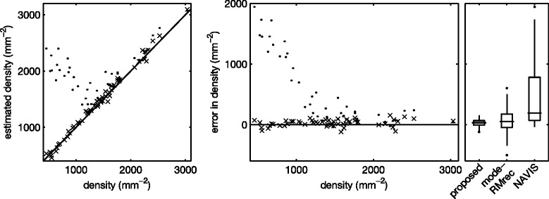
In the first approach, endothelial cell density is estimated with the help of spatial frequency analysis. We evaluate published methods, and propose a new, parameter-free method. In the second approach, based on the stochastic watershed, cells are automatically segmented and the result is used to estimate cell density, polymegathism (cell size variability) and pleomorphism (cell shape variation). We show how to determine optimal values for the three parameters of this algorithm, and compare its results to a semi-automatic delineation by a trained observer.

RESULTS

The frequency analysis method proposed here is more precise than any published method. The segmentation method outperforms the fully automatic method in the NAVIS software (Nidek Technologies Srl, Padova, Italy), which significantly overestimates the number of cells for cell densities below approximately 1200 mm(-2), as well as previously published methods.

CONCLUSIONS

The methods presented here provide a significant improvement over the state of the art, and make in vivo, automated assessment of corneal endothelium more accessible. The segmentation method proposed paves the way to many possible new morphometric parameters, which can quickly and precisely be determined from the segmented image.

摘要

背景

通过共聚焦显微镜在体内获取的图像的手动和半自动分析,常用于确定人眼角膜内皮的质量。这些程序非常耗时。在此,我们提出两种全自动方法来分析和量化通过体内白光裂隙扫描共聚焦显微镜成像的角膜内皮。

方法

在第一种方法中,借助空间频率分析估计内皮细胞密度。我们评估已发表的方法,并提出一种新的、无参数的方法。在第二种方法中,基于随机分水岭算法,自动分割细胞,并将结果用于估计细胞密度、多形性(细胞大小变异性)和异形性(细胞形状变化)。我们展示了如何确定该算法三个参数的最佳值,并将其结果与训练有素的观察者的半自动描绘结果进行比较。

结果

这里提出的频率分析方法比任何已发表的方法都更精确。分割方法优于NAVIS软件(意大利帕多瓦的Nidek Technologies Srl公司)中的全自动方法,该软件对于细胞密度低于约1200 mm⁻² 的情况会显著高估细胞数量,也优于先前发表的方法。

结论

这里提出的方法相较于现有技术有显著改进,使体内角膜内皮的自动评估更容易实现。所提出的分割方法为许多可能的新形态测量参数铺平了道路,这些参数可以从分割图像中快速准确地确定。

https://cdn.ncbi.nlm.nih.gov/pmc/blobs/f257/4451752/118731418343/12880_2015_54_Fig12_HTML.jpg
https://cdn.ncbi.nlm.nih.gov/pmc/blobs/f257/4451752/46a18c152e0b/12880_2015_54_Fig1_HTML.jpg
https://cdn.ncbi.nlm.nih.gov/pmc/blobs/f257/4451752/2388215b1448/12880_2015_54_Fig2_HTML.jpg
https://cdn.ncbi.nlm.nih.gov/pmc/blobs/f257/4451752/5b36c59ae226/12880_2015_54_Fig3_HTML.jpg
https://cdn.ncbi.nlm.nih.gov/pmc/blobs/f257/4451752/29df3dba5f25/12880_2015_54_Fig4_HTML.jpg
https://cdn.ncbi.nlm.nih.gov/pmc/blobs/f257/4451752/3862941eafc9/12880_2015_54_Fig5_HTML.jpg
https://cdn.ncbi.nlm.nih.gov/pmc/blobs/f257/4451752/a71798c4c4d6/12880_2015_54_Fig6_HTML.jpg
https://cdn.ncbi.nlm.nih.gov/pmc/blobs/f257/4451752/03eb0fb5a2a0/12880_2015_54_Fig7_HTML.jpg
https://cdn.ncbi.nlm.nih.gov/pmc/blobs/f257/4451752/0d5f2bc0b0e8/12880_2015_54_Fig8_HTML.jpg
https://cdn.ncbi.nlm.nih.gov/pmc/blobs/f257/4451752/c6598ab97ab8/12880_2015_54_Fig9_HTML.jpg
https://cdn.ncbi.nlm.nih.gov/pmc/blobs/f257/4451752/2bdc5a980873/12880_2015_54_Fig10_HTML.jpg
https://cdn.ncbi.nlm.nih.gov/pmc/blobs/f257/4451752/29c47624059a/12880_2015_54_Fig11_HTML.jpg
https://cdn.ncbi.nlm.nih.gov/pmc/blobs/f257/4451752/118731418343/12880_2015_54_Fig12_HTML.jpg

相似文献

1
Fully automatic evaluation of the corneal endothelium from in vivo confocal microscopy.基于活体共聚焦显微镜的角膜内皮细胞全自动评估
BMC Med Imaging. 2015 Apr 26;15:13. doi: 10.1186/s12880-015-0054-3.
2
A fully automated cell segmentation and morphometric parameter system for quantifying corneal endothelial cell morphology.全自动细胞分割和形态计量参数系统,用于量化角膜内皮细胞形态。
Comput Methods Programs Biomed. 2018 Jul;160:11-23. doi: 10.1016/j.cmpb.2018.03.015. Epub 2018 Mar 22.
3
Dual-model automatic detection of nerve-fibres in corneal confocal microscopy images.角膜共焦显微镜图像中神经纤维的双模型自动检测
Med Image Comput Comput Assist Interv. 2010;13(Pt 1):300-7. doi: 10.1007/978-3-642-15705-9_37.
4
An image model and segmentation algorithm for reflectance confocal images of in vivo cervical tissue.一种用于体内宫颈组织反射共聚焦图像的图像模型与分割算法。
IEEE Trans Image Process. 2005 Sep;14(9):1265-76. doi: 10.1109/tip.2005.852460.
5
Influence of applied corneal endothelium image segmentation techniques on the clinical parameters.应用角膜内皮图像分割技术对临床参数的影响。
Comput Med Imaging Graph. 2017 Jan;55:13-27. doi: 10.1016/j.compmedimag.2016.07.010. Epub 2016 Aug 9.
6
A quantitative comparison between manual segmentation and threshold-based segmentation of CLSM recorded images.共聚焦激光扫描显微镜(CLSM)记录图像的手动分割与基于阈值分割之间的定量比较。
Biomed Sci Instrum. 2007;43:290-5.
7
Development of a Reliable Automated Algorithm for the Morphometric Analysis of Human Corneal Endothelium.用于人角膜内皮形态计量分析的可靠自动化算法的开发
Cornea. 2016 Sep;35(9):1222-8. doi: 10.1097/ICO.0000000000000908.
8
Corneal Endothelial Cell Segmentation by Classifier-Driven Merging of Oversegmented Images.基于分类器的过分割图像融合的角膜内皮细胞分割。
IEEE Trans Med Imaging. 2018 Oct;37(10):2278-2289. doi: 10.1109/TMI.2018.2841910. Epub 2018 May 29.
9
Automatic analysis of diabetic peripheral neuropathy using multi-scale quantitative morphology of nerve fibres in corneal confocal microscopy imaging.使用角膜共聚焦显微镜成像中神经纤维的多尺度定量形态学分析糖尿病周围神经病变。
Med Image Anal. 2011 Oct;15(5):738-47. doi: 10.1016/j.media.2011.05.016. Epub 2011 Jun 13.
10
A reproducible automated segmentation algorithm for corneal epithelium cell images from in vivo laser scanning confocal microscopy.一种从活体激光共聚焦显微镜中获取角膜上皮细胞图像的可重复自动分割算法。
Acta Ophthalmol. 2014 Jun;92(4):e312-6. doi: 10.1111/aos.12304. Epub 2014 Mar 26.

引用本文的文献

1
Open-Source Periorbital Segmentation Dataset for Ophthalmic Applications.用于眼科应用的开源眶周分割数据集。
Ophthalmol Sci. 2025 Mar 5;5(4):100757. doi: 10.1016/j.xops.2025.100757. eCollection 2025 Jul-Aug.
2
Current Applications of Artificial Intelligence for Fuchs Endothelial Corneal Dystrophy: A Systematic Review.人工智能在富克斯内皮性角膜营养不良中的当前应用:一项系统综述。
Transl Vis Sci Technol. 2025 Jun 2;14(6):12. doi: 10.1167/tvst.14.6.12.
3
Data augmentation via warping transforms for modeling natural variability in the corneal endothelium enhances semi-supervised segmentation.

本文引用的文献

1
Assessment of a variable frame (polygonal) method to estimate corneal endothelial cell counts after corneal transplantation.评估一种可变帧(多边形)方法,以估计角膜移植术后的角膜内皮细胞计数。
Eye (Lond). 2012 Jun;26(6):803-9. doi: 10.1038/eye.2012.42. Epub 2012 Mar 23.
2
In vivo confocal microscopy of the corneal endothelium: comparison of three morphometry methods after corneal transplantation.角膜内皮的共聚焦显微镜活体检查:三种角膜移植术后形态计量学方法的比较。
Eye (Lond). 2011 Sep;25(9):1130-7. doi: 10.1038/eye.2011.121. Epub 2011 Jun 10.
3
A system for the automatic estimation of morphometric parameters of corneal endothelium in alizarine red-stained images.
通过变形变换进行数据增强,以模拟角膜内皮的自然变异性,从而增强半监督分割。
PLoS One. 2024 Nov 12;19(11):e0311849. doi: 10.1371/journal.pone.0311849. eCollection 2024.
4
Automatic Determination of Endothelial Cell Density From Donor Cornea Endothelial Cell Images.自动测定供体角膜内皮细胞图像中的内皮细胞密度。
Transl Vis Sci Technol. 2024 Aug 1;13(8):40. doi: 10.1167/tvst.13.8.40.
5
Machine Learning Analysis of Postkeratoplasty Endothelial Cell Images for the Prediction of Future Graft Rejection.基于角膜移植术后内皮细胞图像的机器学习分析预测未来移植物排斥反应。
Transl Vis Sci Technol. 2023 Feb 1;12(2):22. doi: 10.1167/tvst.12.2.22.
6
Corneal endothelium assessment in specular microscopy images with Fuchs' dystrophy via deep regression of signed distance maps.通过带符号距离图的深度回归对伴有Fuchs角膜内皮营养不良的镜面显微镜图像进行角膜内皮评估。
Biomed Opt Express. 2022 Dec 19;14(1):335-351. doi: 10.1364/BOE.477495. eCollection 2023 Jan 1.
7
Mobile-CellNet: Automatic Segmentation of Corneal Endothelium Using an Efficient Hybrid Deep Learning Model.移动细胞网络:使用高效混合深度学习模型自动分割角膜内皮
Cornea. 2023 Apr 1;42(4):456-463. doi: 10.1097/ICO.0000000000003186. Epub 2022 Dec 12.
8
DenseUNets with feedback non-local attention for the segmentation of specular microscopy images of the corneal endothelium with guttae.带有反馈非局部注意力的密集型 UNets 用于分割带胶滴的角膜内皮反射显微镜图像
Sci Rep. 2022 Aug 18;12(1):14035. doi: 10.1038/s41598-022-18180-1.
9
Automated Image Segmentation of the Corneal Endothelium in Patients With Fuchs Dystrophy.自动化图像分割技术在 Fuchs 营养不良患者角膜内皮中的应用。
Transl Vis Sci Technol. 2021 Nov 1;10(13):27. doi: 10.1167/tvst.10.13.27.
10
Advances in Imaging of Subbasal Corneal Nerves With Micro-Optical Coherence Tomography.应用微光学相干断层成像术观察角膜基底部神经的进展
Transl Vis Sci Technol. 2021 Nov 1;10(13):22. doi: 10.1167/tvst.10.13.22.
用于自动估计茜素红染色图像中角膜内皮形态参数的系统。
Br J Ophthalmol. 2010 May;94(5):643-7. doi: 10.1136/bjo.2009.166561.
4
Further analysis of assessments of the coefficient of variation of corneal endothelial cell areas from specular microscopic images.对来自镜面显微镜图像的角膜内皮细胞面积变异系数评估的进一步分析。
Clin Exp Optom. 2008 Sep;91(5):438-46. doi: 10.1111/j.1444-0938.2008.00281.x. Epub 2008 Jun 5.
5
Flat zones filtering, connected operators, and filters by reconstruction.平面区域滤波、连通算子和重建滤波。
IEEE Trans Image Process. 1995;4(8):1153-60. doi: 10.1109/83.403422.
6
Endothelial cell density in donor corneas: a comparison of automatic software programs with manual counting.供体角膜中的内皮细胞密度:自动软件程序与手工计数的比较
Cornea. 2007 Jan;26(1):80-3. doi: 10.1097/ICO.0b013e31802be629.
7
A new system for the automatic estimation of endothelial cell density in donor corneas.一种用于自动估计供体角膜内皮细胞密度的新系统。
Br J Ophthalmol. 2005 Mar;89(3):306-11. doi: 10.1136/bjo.2004.051722.
8
Automatic estimation of endothelium cell density in donor corneas by means of fourier analysis.通过傅里叶分析自动估计供体角膜中的内皮细胞密度。
Med Biol Eng Comput. 2004 Sep;42(5):725-31. doi: 10.1007/BF02347557.
9
Comparison of endothelial cell count using confocal and contact specular microscopy.使用共聚焦显微镜和接触式镜面反射显微镜对内皮细胞计数的比较。
Ophthalmologica. 2003 Mar-Apr;217(2):99-103. doi: 10.1159/000068562.
10
Fourier transform analysis of human corneal endothelial specular photomicrographs.人角膜内皮镜面反射显微镜照片的傅里叶变换分析
Exp Eye Res. 1997 Aug;65(2):205-14. doi: 10.1006/exer.1997.0326.