Krenkel Martin, Markus Andrea, Bartels Matthias, Dullin Christian, Alves Frauke, Salditt Tim
Institute for X-ray Physics, University of Göttingen, 37077 Göttingen, Germany.
Department of Haematology and Medical Oncology, University Medical Center Göttingen, 37075 Göttingen, Germany.
Sci Rep. 2015 May 12;5:9973. doi: 10.1038/srep09973.
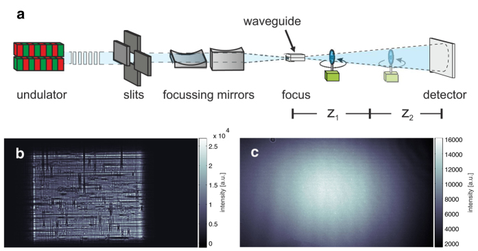
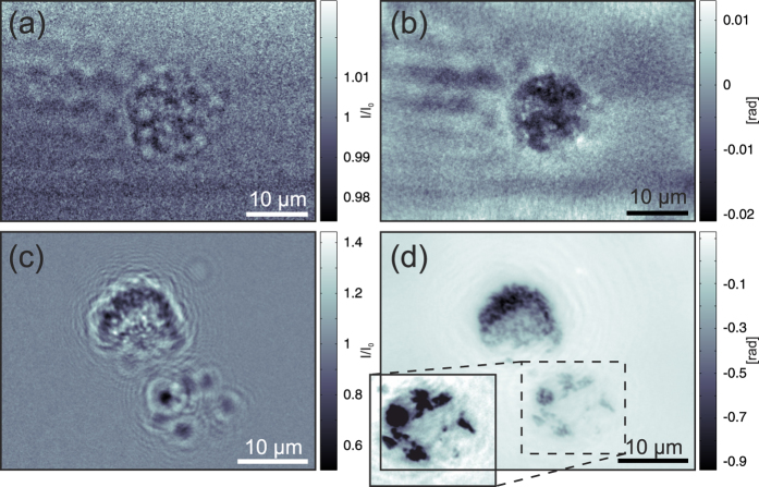
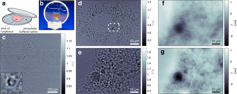
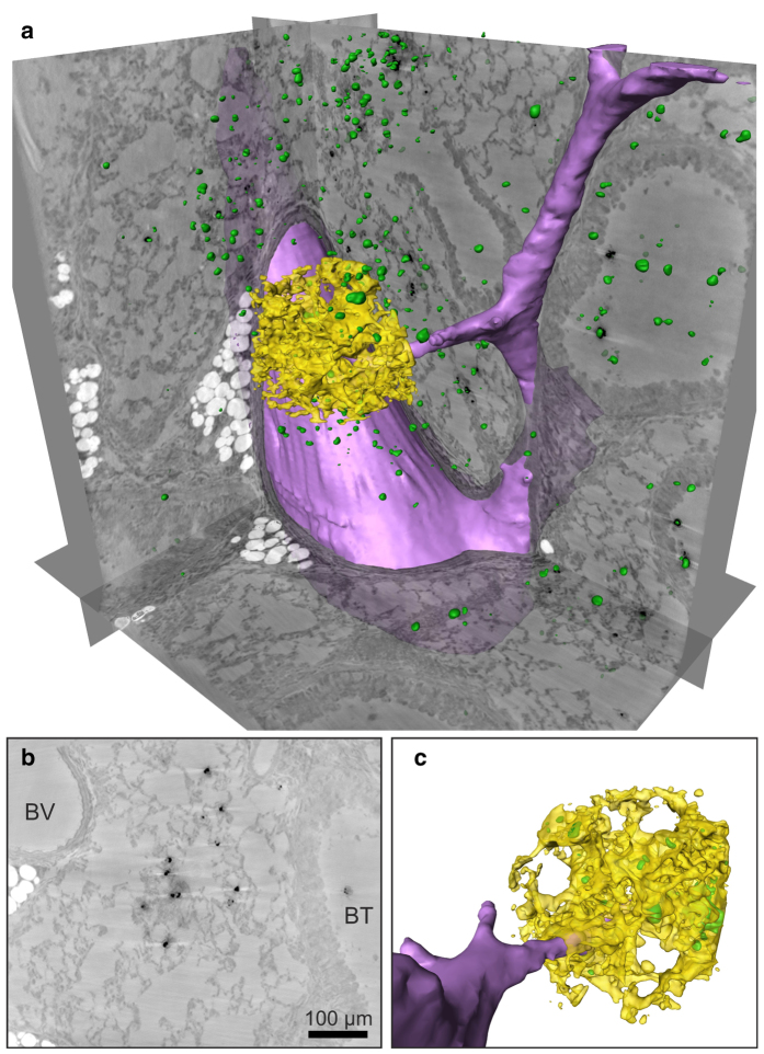
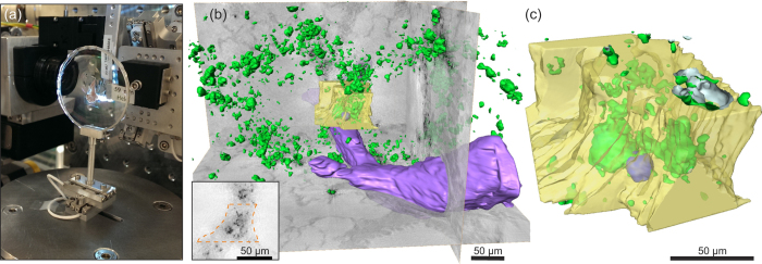
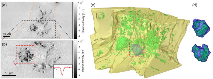
We have performed x-ray phase-contrast tomography on mouse lung tissue. Using a divergent x-ray beam generated by nanoscale focusing, we used zoom tomography to produce three-dimensional reconstructions with selectable magnification, resolution, and field of view. Thus, macroscopic tissue samples extending over several mm can be studied in sub-cellular-level structural detail. The zoom capability and, in particular, the high dose efficiency are enabled by the near-perfect exit wavefront of an optimized x-ray waveguide channel. In combination with suitable phase-retrieval algorithms, challenging radiation-sensitive and low-contrast samples can be reconstructed with minimal artefacts. The dose efficiency of the method is demonstrated by the reconstruction of living macrophages both with and without phagocytized contrast agents. We also used zoom tomography to visualize barium-labelled macrophages in the context of morphological structures in asthmatic and healthy mouse lung tissue one day after intratracheal application. The three-dimensional reconstructions showed that the macrophages predominantly localized to the alveoli, but they were also found in bronchial walls, indicating that these cells might be able to migrate from the lumen of the bronchi through the epithelium.
我们对小鼠肺组织进行了X射线相衬断层扫描。利用纳米级聚焦产生的发散X射线束,我们采用变焦断层扫描技术生成了具有可选择放大倍数、分辨率和视野的三维重建图像。因此,可以在亚细胞水平的结构细节上研究延伸数毫米的宏观组织样本。优化后的X射线波导通道近乎完美的出射波前实现了变焦功能,尤其是高剂量效率。结合合适的相位恢复算法,可以以最小的伪影重建具有挑战性的辐射敏感和低对比度样本。通过对有和没有吞噬造影剂的活巨噬细胞进行重建,证明了该方法的剂量效率。我们还利用变焦断层扫描技术,在气管内给药一天后,观察哮喘和健康小鼠肺组织形态结构背景下钡标记的巨噬细胞。三维重建显示,巨噬细胞主要定位于肺泡,但也存在于支气管壁,这表明这些细胞可能能够从支气管腔穿过上皮迁移。