Klöppel Stefan, Peter Jessica, Ludl Anna, Pilatus Anne, Maier Sabrina, Mader Irina, Heimbach Bernhard, Frings Lars, Egger Karl, Dukart Juergen, Schroeter Matthias L, Perneczky Robert, Häussermann Peter, Vach Werner, Urbach Horst, Teipel Stefan, Hüll Michael, Abdulkadir Ahmed
Center of Geriatrics and Gerontology Freiburg, University Medical Center Freiburg, Freiburg, Germany.
Freiburg Brain Imaging, University Medical Center Freiburg, Germany.
J Alzheimers Dis. 2015;47(4):939-54. doi: 10.3233/JAD-150334.
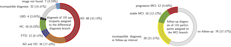
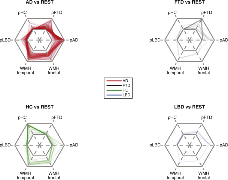
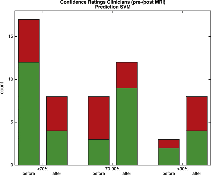
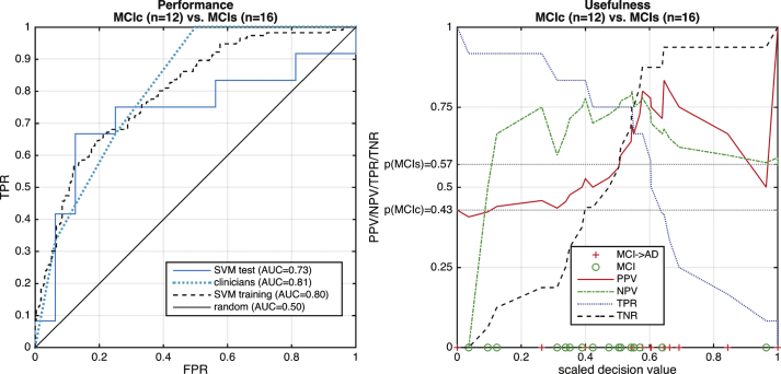
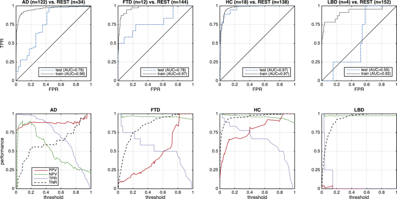
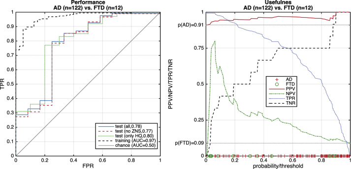
Several studies have demonstrated that fully automated pattern recognition methods applied to structural magnetic resonance imaging (MRI) aid in the diagnosis of dementia, but these conclusions are based on highly preselected samples that significantly differ from that seen in a dementia clinic. At a single dementia clinic, we evaluated the ability of a linear support vector machine trained with completely unrelated data to differentiate between Alzheimer's disease (AD), frontotemporal dementia (FTD), Lewy body dementia, and healthy aging based on 3D-T1 weighted MRI data sets. Furthermore, we predicted progression to AD in subjects with mild cognitive impairment (MCI) at baseline and automatically quantified white matter hyperintensities from FLAIR-images. Separating additionally recruited healthy elderly from those with dementia was accurate with an area under the curve (AUC) of 0.97 (according to Fig. 4). Multi-class separation of patients with either AD or FTD from other included groups was good on the training set (AUC > 0.9) but substantially less accurate (AUC = 0.76 for AD, AUC = 0.78 for FTD) on 134 cases from the local clinic. Longitudinal data from 28 cases with MCI at baseline and appropriate follow-up data were available. The computer tool discriminated progressive from stable MCI with AUC = 0.73, compared to AUC = 0.80 for the training set. A relatively low accuracy by clinicians (AUC = 0.81) illustrates the difficulties of predicting conversion in this heterogeneous cohort. This first application of a MRI-based pattern recognition method to a routine sample demonstrates feasibility, but also illustrates that automated multi-class differential diagnoses have to be the focus of future methodological developments and application studies.
多项研究表明,应用于结构磁共振成像(MRI)的全自动模式识别方法有助于痴呆症的诊断,但这些结论是基于高度预选的样本得出的,这些样本与痴呆症诊所中所见的样本有显著差异。在一家单一的痴呆症诊所,我们评估了一种用完全不相关数据训练的线性支持向量机,基于三维T1加权MRI数据集区分阿尔茨海默病(AD)、额颞叶痴呆(FTD)、路易体痴呆和健康衰老的能力。此外,我们预测了基线时轻度认知障碍(MCI)患者进展为AD的情况,并从液体衰减反转恢复(FLAIR)图像中自动量化了白质高信号。将额外招募的健康老年人与痴呆症患者区分开来的准确率较高,曲线下面积(AUC)为0.97(根据图4)。在训练集上,将AD或FTD患者与其他纳入组进行多类别区分的效果良好(AUC>0.9),但在当地诊所的134例病例中,准确率大幅降低(AD的AUC = 0.76,FTD的AUC = 0.78)。有来自28例基线时患有MCI且有适当随访数据的纵向数据。与训练集的AUC = 0.80相比,该计算机工具区分进展性MCI和稳定MCI的AUC = 0.73。临床医生的准确率相对较低(AUC = 0.81),这说明了在这个异质性队列中预测转化的困难。这种基于MRI的模式识别方法在常规样本中的首次应用证明了其可行性,但也表明自动化多类别鉴别诊断必须成为未来方法学发展和应用研究的重点。