Raman Rajiv, Nittala Muneeswar Gupta, Gella Laxmi, Pal Swakshyar Saumya, Sharma Tarun
Shri Bhagwan Mahavir Vitreoretinal Services, Sankara Nethralaya, Chennai, Tamil Nadu, India.
Department of Optometry, Elite School of Optometry, Chennai, Tamil Nadu, India.
J Ophthalmic Vis Res. 2015 Apr-Jun;10(2):160-4. doi: 10.4103/2008-322X.163771.
To evaluate retinal sensitivity over hard exudates in correlation with the spectral domain optical coherence tomography (SD-OCT) findings in eyes with diabetic retinopathy.
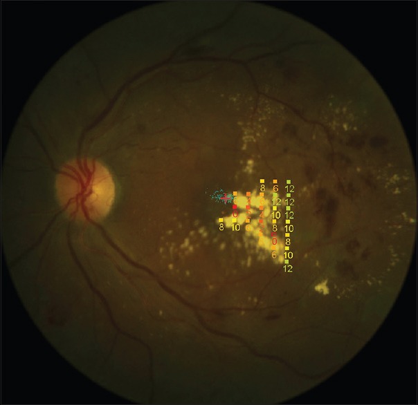
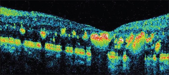
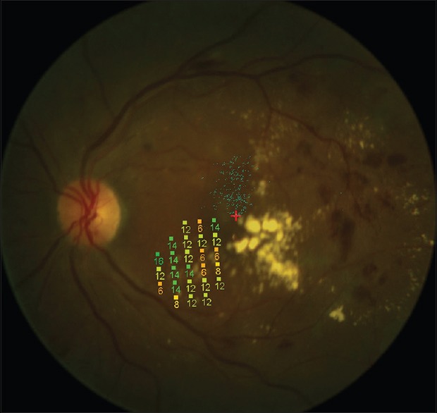
Twelve eyes of 10 patients with hard exudates associated with diabetic retinopathy were enrolled in this study. All subjects underwent a complete ophthalmic examination including SD-OCT (Copernicus, Zawiercie, Poland) and microperimetry (MP1; Nidek Technologies, Padova, Italy). Retinal sensitivity was measured, over the areas with hard exudates and compared to corresponding locations devoid of hard exudates, using a semi-automatic program. The size of the hard exudate plaque was measured using the measurement software in the microperimeter. Retinal thickness in the area of the hard exudates and foveal thickness were measured using SD-OCT.
Mean retinal sensitivity over hard exudates was 4.97 ± 4.17 dB which was significantly (P = 0.0001) reduced as compared to locations devoid of hard exudates. No significant correlation (r=-0.23, P = 0.45) was found between the size of the hard exudates and retinal sensitivity. A significant negative correlation was found between retinal sensitivity and retinal thickness at the area of the hard exudates (r=-0.65, P = 0.05), and between retinal sensitivity and foveal thickness (r=-0.91, P = 0.001).
In eyes with diabetic retinopathy, retinal sensitivity was reduced due to the presence of hard exudates in the outer retinal layers and retinal thickening but this was not correlated with the size of the hard exudates.
评估糖尿病视网膜病变患者眼中硬性渗出物上方的视网膜敏感度,并将其与光谱域光学相干断层扫描(SD-OCT)结果相关联。
本研究纳入了10例患有与糖尿病视网膜病变相关的硬性渗出物的患者的12只眼睛。所有受试者均接受了包括SD-OCT(哥白尼,扎维尔切,波兰)和微视野计(MP1;尼德克科技公司,帕多瓦,意大利)在内的全面眼科检查。使用半自动程序测量硬性渗出物区域的视网膜敏感度,并与没有硬性渗出物的相应位置进行比较。使用微视野计中的测量软件测量硬性渗出斑块的大小。使用SD-OCT测量硬性渗出物区域的视网膜厚度和黄斑中心凹厚度。
硬性渗出物上方的平均视网膜敏感度为4.97±4.17 dB,与没有硬性渗出物的位置相比,显著降低(P = 0.0001)。硬性渗出物的大小与视网膜敏感度之间未发现显著相关性(r = -0.23,P = 0.45)。在硬性渗出物区域,视网膜敏感度与视网膜厚度之间存在显著负相关(r = -0.65,P = 0.05),在视网膜敏感度与黄斑中心凹厚度之间也存在显著负相关(r = -0.91,P = 0.001)。
在糖尿病视网膜病变患者的眼中,由于视网膜外层存在硬性渗出物和视网膜增厚,视网膜敏感度降低,但这与硬性渗出物的大小无关。