• 文献检索
  • 文档翻译
  • 深度研究
  • 学术资讯
  • Suppr Zotero 插件Zotero 插件
  • 邀请有礼
  • 套餐&价格
  • 历史记录
应用&插件
Suppr Zotero 插件Zotero 插件浏览器插件Mac 客户端Windows 客户端微信小程序
定价
高级版会员购买积分包购买API积分包
服务
文献检索文档翻译深度研究API 文档MCP 服务
关于我们
关于 Suppr公司介绍联系我们用户协议隐私条款
关注我们

Suppr 超能文献

核心技术专利:CN118964589B侵权必究
粤ICP备2023148730 号-1Suppr @ 2026

文献检索

告别复杂PubMed语法,用中文像聊天一样搜索,搜遍4000万医学文献。AI智能推荐,让科研检索更轻松。

立即免费搜索

文件翻译

保留排版,准确专业,支持PDF/Word/PPT等文件格式,支持 12+语言互译。

免费翻译文档

深度研究

AI帮你快速写综述,25分钟生成高质量综述,智能提取关键信息,辅助科研写作。

立即免费体验

三种量子点(QDs)的阈剂量在小鼠成纤维细胞L929中诱导氧化应激,引发DNA损伤和细胞凋亡。

Threshold Dose of Three Types of Quantum Dots (QDs) Induces Oxidative Stress Triggers DNA Damage and Apoptosis in Mouse Fibroblast L929 Cells.

作者信息

Zhang Ting, Wang Yiqing, Kong Lu, Xue Yuying, Tang Meng

机构信息

Key Laboratory of Environmental Medicine Engineering, Ministry of Education, School of Public Health, Southeast University, Nanjing 210009, China.

Jiangsu Key Laboratory for Biomaterials and Devices, Southeast University, Nanjing 210009, China.

出版信息

Int J Environ Res Public Health. 2015 Oct 26;12(10):13435-54. doi: 10.3390/ijerph121013435.

DOI:10.3390/ijerph121013435
PMID:26516873
原文链接:https://pmc.ncbi.nlm.nih.gov/articles/PMC4627041/
Abstract

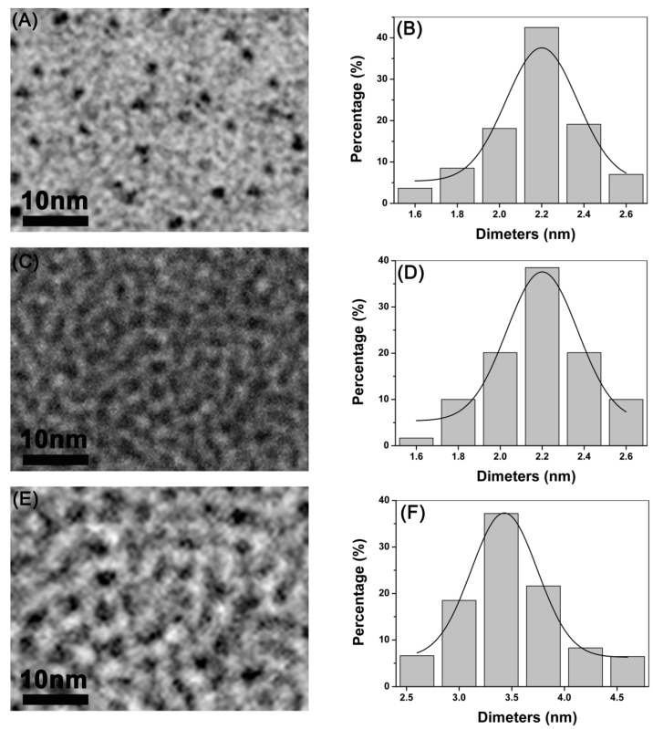
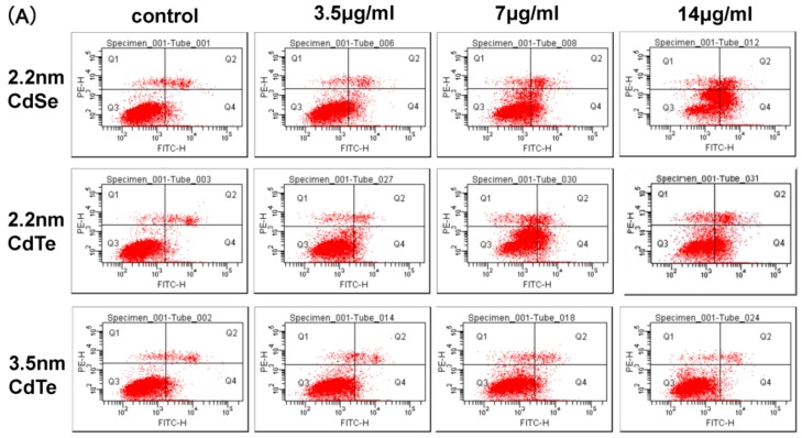
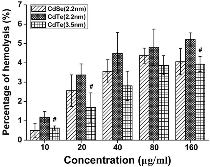
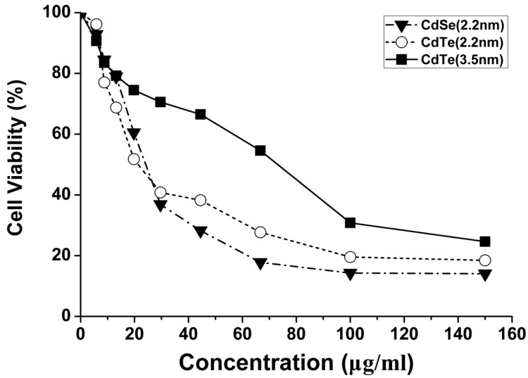
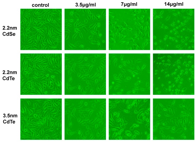
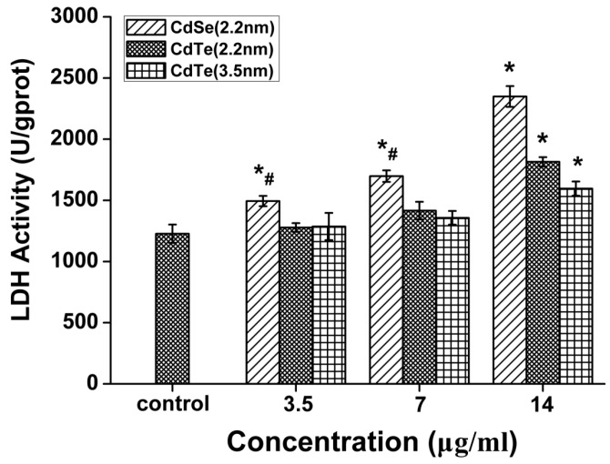
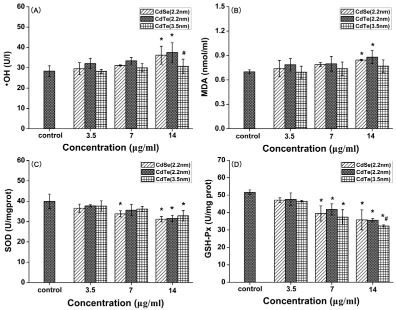
Although it has been reported that fluorescent quantum dots (QDs) have obvious acute toxic effects in vitro, their toxic effects at low doses or threshold doses are still unknown. Therefore, we evaluated the biological histocompatibility and in vitro toxicity of three types of QDs at threshold doses. Also, we compared the toxic effects of QDs with different raw chemical compositions and sizes. The results showed that low concentrations of QDs (≤7 μg/mL) had no obvious effect on cell viability and cell membrane damage, oxidative damage, cell apoptosis or DNA damage. However, QD exposure led to a significant cytotoxicity at higher doses (≥14 μg/mL) and induced abnormal cellular morphology. In addition, when comparing the three types of QDs, 2.2 nm CdTe QDs exposure showed a significantly increased proportion of apoptotic cells and significant DNA damage, suggesting that size and composition contribute to the toxic effects of QDs. Based on these discussions, it was concluded that the concentration (7 μg/mL) may serve as a threshold level for these three types of QDs only in L929 fibroblasts, whereas high concentrations (above 14 μg/mL) may be toxic, resulting in inhibition of proliferation, induction of apoptosis and DNA damage in L929 fibroblasts.

摘要

尽管有报道称荧光量子点(QDs)在体外具有明显的急性毒性作用,但其在低剂量或阈剂量下的毒性作用仍不清楚。因此,我们评估了三种类型的量子点在阈剂量下的生物组织相容性和体外毒性。此外,我们比较了具有不同原始化学成分和尺寸的量子点的毒性作用。结果表明,低浓度的量子点(≤7μg/mL)对细胞活力、细胞膜损伤、氧化损伤、细胞凋亡或DNA损伤没有明显影响。然而,在较高剂量(≥14μg/mL)下,量子点暴露会导致显著的细胞毒性并诱导细胞形态异常。此外,在比较这三种类型的量子点时,暴露于2.2nm的碲化镉量子点显示凋亡细胞比例显著增加且DNA损伤显著,这表明尺寸和成分会影响量子点的毒性作用。基于这些讨论,得出的结论是,浓度(7μg/mL)可能仅在L929成纤维细胞中作为这三种类型量子点的阈水平,而高浓度(高于14μg/mL)可能具有毒性,导致L929成纤维细胞增殖受抑制、诱导凋亡和DNA损伤。

https://cdn.ncbi.nlm.nih.gov/pmc/blobs/49b5/4627041/ac60987d6b76/ijerph-12-13435-g007a.jpg
https://cdn.ncbi.nlm.nih.gov/pmc/blobs/49b5/4627041/35d4da261c08/ijerph-12-13435-g001.jpg
https://cdn.ncbi.nlm.nih.gov/pmc/blobs/49b5/4627041/ea71946af54b/ijerph-12-13435-g002.jpg
https://cdn.ncbi.nlm.nih.gov/pmc/blobs/49b5/4627041/b69af43c80ae/ijerph-12-13435-g003.jpg
https://cdn.ncbi.nlm.nih.gov/pmc/blobs/49b5/4627041/fd4befe43b2f/ijerph-12-13435-g004.jpg
https://cdn.ncbi.nlm.nih.gov/pmc/blobs/49b5/4627041/5d3abd724451/ijerph-12-13435-g005.jpg
https://cdn.ncbi.nlm.nih.gov/pmc/blobs/49b5/4627041/9abec39e3430/ijerph-12-13435-g006.jpg
https://cdn.ncbi.nlm.nih.gov/pmc/blobs/49b5/4627041/ac60987d6b76/ijerph-12-13435-g007a.jpg

相似文献

1
Threshold Dose of Three Types of Quantum Dots (QDs) Induces Oxidative Stress Triggers DNA Damage and Apoptosis in Mouse Fibroblast L929 Cells.三种量子点(QDs)的阈剂量在小鼠成纤维细胞L929中诱导氧化应激,引发DNA损伤和细胞凋亡。
Int J Environ Res Public Health. 2015 Oct 26;12(10):13435-54. doi: 10.3390/ijerph121013435.
2
Dose- and duration-dependent cytotoxicity and genotoxicity in human hepato carcinoma cells due to CdTe QDs exposure.CdTe QDs 暴露导致人肝癌细胞的剂量和时间依赖性细胞毒性和遗传毒性。
Hum Exp Toxicol. 2019 Aug;38(8):914-926. doi: 10.1177/0960327119843578. Epub 2019 Apr 17.
3
Toxicity assessment of repeated intravenous injections of arginine-glycine-aspartic acid peptide conjugated CdSeTe/ZnS quantum dots in mice.小鼠重复静脉注射精氨酸-甘氨酸-天冬氨酸肽偶联的CdSeTe/ZnS量子点的毒性评估
Int J Nanomedicine. 2014 Oct 17;9:4809-17. doi: 10.2147/IJN.S70092. eCollection 2014.
4
Cytotoxicity assessment of functionalized CdSe, CdTe and InP quantum dots in two human cancer cell models.功能化 CdSe、CdTe 和 InP 量子点在两种人类癌细胞模型中的细胞毒性评估。
Mater Sci Eng C Mater Biol Appl. 2015 Dec 1;57:222-31. doi: 10.1016/j.msec.2015.07.044. Epub 2015 Jul 26.
5
[Oxidative damage effects induced by CdTe quantum dots in mice].碲化镉量子点对小鼠的氧化损伤效应
Zhonghua Lao Dong Wei Sheng Zhi Ye Bing Za Zhi. 2017 Jul 20;35(7):487-490. doi: 10.3760/cma.j.issn.1001-9391.2017.07.003.
6
[Oxidative damage effects induced by CdTe quantum dots in mice liver].碲化镉量子点对小鼠肝脏的氧化损伤作用
Wei Sheng Yan Jiu. 2013 Jan;42(1):39-43.
7
Determination of a threshold dose to reduce or eliminate CdTe-induced toxicity in L929 cells by controlling the exposure dose.通过控制暴露剂量,确定降低或消除 CdTe 诱导的 L929 细胞毒性的阈值剂量。
PLoS One. 2013;8(4):e59359. doi: 10.1371/journal.pone.0059359. Epub 2013 Apr 5.
8
The protective effects of resveratrol, HS and thermotherapy on the cell apoptosis induced by CdTe quantum dots.白藜芦醇、硫化氢和热疗对碲化镉量子点诱导的细胞凋亡的保护作用。
Toxicol In Vitro. 2017 Jun;41:106-113. doi: 10.1016/j.tiv.2017.02.013. Epub 2017 Feb 20.
9
Acute and chronic cadmium telluride quantum dots-exposed human bronchial epithelial cells: The effects of particle sizes on their cytotoxicity and carcinogenicity.急性和慢性暴露于碲化镉量子点的人支气管上皮细胞:颗粒大小对其细胞毒性和致癌性的影响。
Biochem Biophys Res Commun. 2018 Jan 1;495(1):899-903. doi: 10.1016/j.bbrc.2017.11.074. Epub 2017 Nov 12.
10
Conformational and functional effects of MPA-CdTe quantum dots on SOD: Evaluating the mechanism of oxidative stress induced by quantum dots in the mouse nephrocytes.MPA-CdTe 量子点对 SOD 的构象和功能影响:评估量子点在小鼠肾细胞中诱导氧化应激的机制。
J Mol Recognit. 2019 Sep;32(9):e2783. doi: 10.1002/jmr.2783. Epub 2019 May 2.

引用本文的文献

1
A Review of in vivo Toxicity of Quantum Dots in Animal Models.量子点在动物模型体内毒性的综述。
Int J Nanomedicine. 2023 Dec 29;18:8143-8168. doi: 10.2147/IJN.S434842. eCollection 2023.
2
Feasibility of Silicon Quantum Dots as a Biomarker for the Bioimaging of Tear Film.硅量子点作为泪膜生物成像生物标志物的可行性
Nanomaterials (Basel). 2022 Jun 8;12(12):1965. doi: 10.3390/nano12121965.
3
Assessment of the Toxicity of Quantum Dots through Biliometric Analysis.通过比色法分析评估量子点的毒性

本文引用的文献

1
Probing the Cytotoxicity Of Semiconductor Quantum Dots.探究半导体量子点的细胞毒性
Nano Lett. 2004 Jan 1;4(1):11-18. doi: 10.1021/nl0347334. Epub 2003 Dec 10.
2
Liver Toxicity of Cadmium Telluride Quantum Dots (CdTe QDs) Due to Oxidative Stress in Vitro and in Vivo.碲化镉量子点(CdTe QDs)在体外和体内因氧化应激导致的肝脏毒性
Int J Mol Sci. 2015 Sep 25;16(10):23279-99. doi: 10.3390/ijms161023279.
3
Mechanistic Investigation of the Biological Effects of SiO₂, TiO₂, and ZnO Nanoparticles on Intestinal Cells.SiO₂、TiO₂ 和 ZnO 纳米颗粒对肠道细胞的生物学效应的机制研究。
Int J Environ Res Public Health. 2021 May 27;18(11):5768. doi: 10.3390/ijerph18115768.
4
Comparing Transcriptome Profiles of Saccharomyces Cerevisiae Cells Exposed to Cadmium Selenide/Zinc Sulfide and Indium Phosphide/Zinc Sulfide.比较暴露于碲化镉/硫化锌和磷化铟/硫化锌的酿酒酵母细胞的转录组图谱。
Genes (Basel). 2021 Mar 17;12(3):428. doi: 10.3390/genes12030428.
5
CdSe/ZnS Core-Shell-Type Quantum Dot Nanoparticles Disrupt the Cellular Homeostasis in Cellular Blood-Brain Barrier Models.硒化镉/硫化锌核壳型量子点纳米颗粒破坏细胞血脑屏障模型中的细胞内稳态。
Int J Mol Sci. 2021 Jan 22;22(3):1068. doi: 10.3390/ijms22031068.
6
Quantum Dots: An Emerging Tool for Point-of-Care Testing.量子点:即时检验的新兴工具。
Micromachines (Basel). 2020 Nov 29;11(12):1058. doi: 10.3390/mi11121058.
7
Mixed Mercaptocarboxylic Acid Shells Provide Stable Dispersions of InPZnS/ZnSe/ZnS Multishell Quantum Dots in Aqueous Media.混合巯基羧酸壳层使InPZnS/ZnSe/ZnS多壳层量子点在水介质中具有稳定的分散性。
Nanomaterials (Basel). 2020 Sep 17;10(9):1858. doi: 10.3390/nano10091858.
8
Cadmium telluride quantum dot-exposed human bronchial epithelial cells: a further study of the cellular response by proteomics.碲化镉量子点暴露的人支气管上皮细胞:通过蛋白质组学对细胞反应的进一步研究
Toxicol Res (Camb). 2019 Oct 22;8(6):994-1001. doi: 10.1039/c9tx00126c. eCollection 2019 Nov 1.
9
Revisiting the cytotoxicity of quantum dots: an in-depth overview.重新审视量子点的细胞毒性:深入概述。
Biophys Rev. 2020 Jun;12(3):703-718. doi: 10.1007/s12551-020-00653-0. Epub 2020 Mar 5.
10
ToxTracker Reporter Cell Lines as a Tool for Mechanism-Based (geno)Toxicity Screening of Nanoparticles-Metals, Oxides and Quantum Dots.ToxTracker报告细胞系作为基于机制的纳米颗粒(金属、氧化物和量子点)(基因)毒性筛选工具
Nanomaterials (Basel). 2020 Jan 6;10(1):110. doi: 10.3390/nano10010110.
Small. 2015 Jul;11(28):3458-68. doi: 10.1002/smll.201403232. Epub 2015 Apr 22.
4
Understanding nanoparticle cellular entry: A physicochemical perspective.理解纳米颗粒的细胞内进入:物理化学视角。
Adv Colloid Interface Sci. 2015 Apr;218:48-68. doi: 10.1016/j.cis.2015.01.007. Epub 2015 Feb 12.
5
Molybdenum nanoparticles-induced cytotoxicity, oxidative stress, G2/M arrest, and DNA damage in mouse skin fibroblast cells (L929).钼纳米颗粒诱导的小鼠皮肤成纤维细胞(L929)的细胞毒性、氧化应激、G2/M 期阻滞和 DNA 损伤。
Colloids Surf B Biointerfaces. 2015 Jan 1;125:73-81. doi: 10.1016/j.colsurfb.2014.11.014. Epub 2014 Nov 20.
6
Genotoxic effects of CdS quantum dots and Ag2S nanoparticles in fish cell lines (RTG-2).硫化镉量子点和硫化银纳米颗粒对鱼类细胞系(RTG-2)的遗传毒性作用。
Mutat Res Genet Toxicol Environ Mutagen. 2014 Dec;775-776:89-93. doi: 10.1016/j.mrgentox.2014.09.003. Epub 2014 Sep 16.
7
Quantum dots-bevacizumab complexes for in vivo imaging of tumors.用于肿瘤体内成像的量子点-贝伐单抗复合物
In Vivo. 2014 Nov-Dec;28(6):1091-5.
8
Cytotoxicity, DNA damage, and apoptosis induced by titanium dioxide nanoparticles in human non-small cell lung cancer A549 cells.二氧化钛纳米颗粒对人非小细胞肺癌A549细胞的细胞毒性、DNA损伤及凋亡作用
Environ Sci Pollut Res Int. 2015 Apr;22(7):5519-30. doi: 10.1007/s11356-014-3717-7. Epub 2014 Oct 24.
9
DNA damage and oxidative stress induced by CeO2 nanoparticles in human dermal fibroblasts: Evidence of a clastogenic effect as a mechanism of genotoxicity.CeO2 纳米颗粒在人真皮成纤维细胞中诱导的 DNA 损伤和氧化应激:作为遗传毒性机制的断裂作用证据。
Nanotoxicology. 2015;9(6):696-705. doi: 10.3109/17435390.2014.968889. Epub 2014 Oct 17.
10
Nonlinear effects of nanoparticles: biological variability from hormetic doses, small particle sizes, and dynamic adaptive interactions.纳米粒子的非线性效应:从兴奋剂量、小颗粒尺寸和动态自适应相互作用方面看生物变异性。
Dose Response. 2013 Nov 7;12(2):202-32. doi: 10.2203/dose-response.13-025.Bell. eCollection 2014 May.