Monari Emanuela, Cuoghi Aurora, Bellei Elisa, Bergamini Stefania, Lucchi Andrea, Tomasi Aldo, Cortellini Pierpaolo, Zaffe Davide, Bertoldi Carlo
Department of Diagnostic, Clinical and Public Health Medicine, University of Modena and Reggio Emilia, Largo del Pozzo, 71-41124 Modena, Italy.
Private Practice, Modena, Italy.
Proteome Sci. 2015 Dec 30;13:33. doi: 10.1186/s12953-015-0089-y. eCollection 2015.
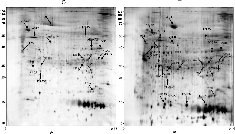
The periodontal disease is caused by a set of inflammatory disorders characterized by periodontal pocket formation that lead to tooth loss if untreated. The proteomic profile and related molecular conditions of pocket tissue in periodontally-affected patients are not reported in literature. To characterize the proteomic profile of periodontally-affected patients, their interproximal periodontal pocket tissue was compared with that of periodontally-healthy patients. Pocket-associated and healthy tissue samples, harvested during surgical therapy, were treated to extract the protein content. Tissues were always collected at sites where no periodontal-pathogenic bacteria were detectable. Proteins were separated using two-dimensional gel electrophoresis and identified by liquid chromatography/mass spectrometry. After identification, four proteins were selected for subsequent Western Blot quantitation both in pathological and healty tissues.
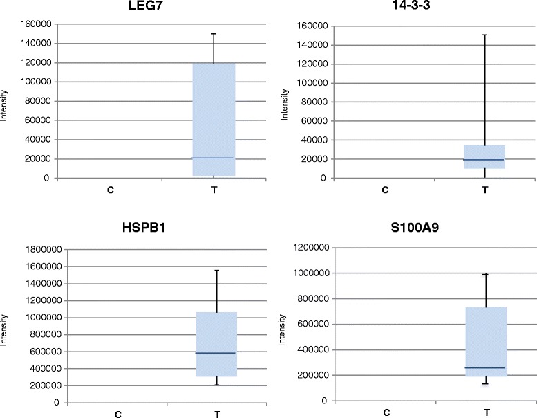
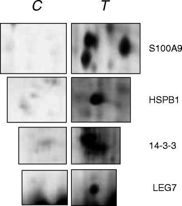
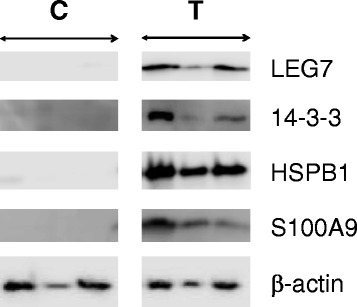
A significant unbalance in protein expression between healthy and pathological sites was recorded. Thirty-two protein spots were overall identified, and four proteins (S100A9, HSPB1, LEG7 and 14-3-3) were selected for Western blot analysis of both periodontally-affected and healthy patients. The four selected proteins resulted over-expressed in periodontal pocket tissue when compared with the corresponding tissue of periodontally-healthy patients. The results of Western blot analysis are congruent with the defensive and the regenerative reaction of injured periodontal tissues.
The proteomic analysis was performed for the first time directly on periodontal pocket tissue. The proteomic network highlighted in this study enhances the understanding of periodontal disease pathogenesis necessary for specific therapeutic strategies setting.
牙周病是由一系列炎症性疾病引起的,其特征为牙周袋形成,若不治疗会导致牙齿脱落。牙周病患者牙周袋组织的蛋白质组学特征及相关分子状况在文献中未见报道。为了表征牙周病患者的蛋白质组学特征,将其邻面牙周袋组织与牙周健康患者的组织进行了比较。在手术治疗期间采集的牙周袋相关组织和健康组织样本,经过处理以提取蛋白质含量。组织总是在检测不到牙周病原菌的部位采集。蛋白质通过二维凝胶电泳进行分离,并通过液相色谱/质谱进行鉴定。鉴定后,选择了四种蛋白质在病理组织和健康组织中进行后续的蛋白质印迹定量分析。
记录到健康部位和病理部位之间蛋白质表达存在显著失衡。总共鉴定出32个蛋白质斑点,并选择了四种蛋白质(S100A9、HSPB1、LEG7和14-3-3)对牙周病患者和健康患者进行蛋白质印迹分析。与牙周健康患者的相应组织相比,所选的四种蛋白质在牙周袋组织中过度表达。蛋白质印迹分析结果与受损牙周组织的防御和再生反应一致。
首次直接对牙周袋组织进行蛋白质组学分析。本研究中突出显示的蛋白质组学网络增强了对制定特定治疗策略所必需的牙周病发病机制的理解。