Brain Research Imaging Centre, The University of Edinburgh, Edinburgh EH4 2XU, UK.
Centre for Clinical Brain Sciences, The University of Edinburgh, Edinburgh EH16 4SB, UK.
Sci Data. 2016 Feb 2;3:160003. doi: 10.1038/sdata.2016.3.
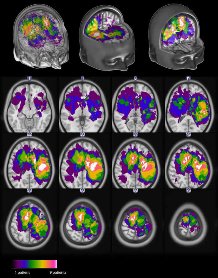
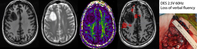
We collected high resolution structural (T1, T2, DWI) and several functional (BOLD T2*) MRI data in 22 patients with different types of brain tumours. Functional imaging protocols included a motor task, a verb generation task, a word repetition task and resting state. Imaging data are complemented by demographics (age, sex, handedness, and pathology), behavioural results to motor and cognitive tests and direct cortical electrical stimulation data (pictures of stimulation sites with outcomes) performed during surgery. Altogether, these data are suited to test functional imaging methods for single subject analyses, in particular methods that focus on locating eloquent cortical areas, critical functional and/or structural network hubs, and predict patient status based on imaging data (presurgical mapping).
我们收集了 22 名患有不同类型脑肿瘤患者的高分辨率结构(T1、T2、DWI)和多种功能(BOLD T2*)MRI 数据。功能成像方案包括运动任务、动词生成任务、单词重复任务和静息状态。成像数据辅以人口统计学信息(年龄、性别、利手性和病理学)、运动和认知测试的行为结果以及手术期间进行的直接皮质电刺激数据(带有结果的刺激部位图片)。总之,这些数据适合用于测试针对单个体分析的功能成像方法,特别是那些专注于定位语言相关皮质区、关键功能和/或结构网络枢纽以及基于成像数据预测患者状态(术前映射)的方法。