Wei Yan-Yan, Wang Ji-Jun, Yan Chao, Li Zi-Qiang, Pan Xiao, Cui Yi, Su Tong, Liu Tao-Sheng, Tang Yun-Xiang
Department of Medical Psychology, Faculty of Mental Health, Second Military Medical University, Shanghai 200433, China.
Chin Med J (Engl). 2016 Mar 5;129(5):578-85. doi: 10.4103/0366-6999.176983.
Several studies using functional magnetic resonance imaging (fMRI) and positron emission tomography (PET) have indicated that cognitive remediation therapy (CRT) might improve cognitive function by changing brain activations in patients with schizophrenia. However, the results were not consistent in these changed brain areas in different studies. The present activation likelihood estimation (ALE) meta-analysis was conducted to investigate whether cognitive function change was accompanied by the brain activation changes, and where the main areas most related to these changes were in schizophrenia patients after CRT. Analyses of whole-brain studies and whole-brain + region of interest (ROI) studies were compared to explore the effect of the different methodologies on the results.
A computerized systematic search was conducted to collect fMRI and PET studies on brain activation changes in schizophrenia patients from pre- to post-CRT. Nine studies using fMRI techniques were included in the meta-analysis. Ginger ALE 2.3.1 was used to perform meta-analysis across these imaging studies.
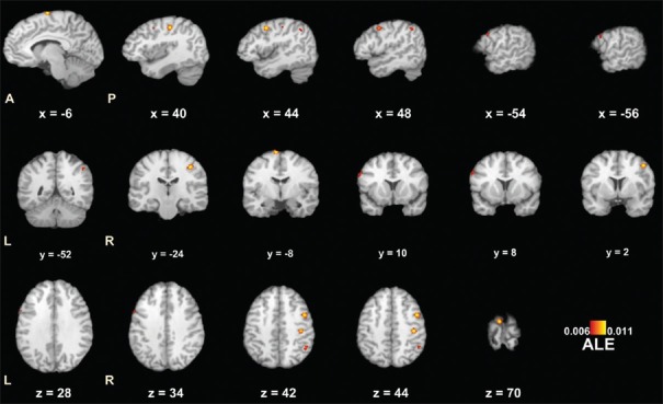
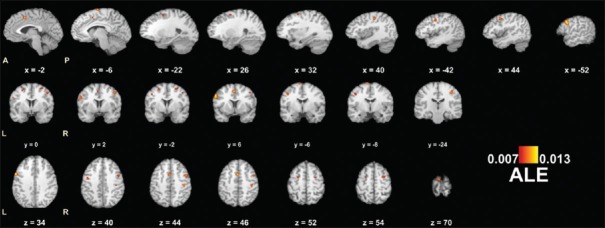
The main areas with increased brain activation were in frontal and parietal lobe, including left medial frontal gyrus, left inferior frontal gyrus, right middle frontal gyrus, right postcentral gyrus, and inferior parietal lobule in patients after CRT, yet no decreased brain activation was found. Although similar increased activation brain areas were identified in ALE with or without ROI studies, analysis including ROI studies had a higher ALE value.
The current findings suggest that CRT might improve the cognition of schizophrenia patients by increasing activations of the frontal and parietal lobe. In addition, it might provide more evidence to confirm results by including ROI studies in ALE meta-analysis.
多项使用功能磁共振成像(fMRI)和正电子发射断层扫描(PET)的研究表明,认知康复疗法(CRT)可能通过改变精神分裂症患者的脑激活来改善认知功能。然而,在不同研究中,这些脑区变化的结果并不一致。本研究进行激活可能性估计(ALE)元分析,以调查认知功能变化是否伴随着脑激活变化,以及CRT后精神分裂症患者中与这些变化最相关的主要区域在哪里。比较了全脑研究和全脑+感兴趣区域(ROI)研究的分析,以探讨不同方法对结果的影响。
进行计算机系统检索,以收集关于精神分裂症患者CRT前后脑激活变化的fMRI和PET研究。元分析纳入了9项使用fMRI技术的研究。使用Ginger ALE 2.3.1对这些影像学研究进行元分析。
CRT后患者脑激活增加的主要区域位于额叶和顶叶,包括左侧额内侧回、左侧额下回、右侧额中回、右侧中央后回和顶下小叶,未发现脑激活减少。尽管在有无ROI研究的ALE中都发现了类似的脑激活增加区域,但包括ROI研究的分析具有更高的ALE值。
目前的研究结果表明,CRT可能通过增加额叶和顶叶的激活来改善精神分裂症患者的认知。此外,在ALE元分析中纳入ROI研究可能为证实结果提供更多证据。