• 文献检索
  • 文档翻译
  • 深度研究
  • 学术资讯
  • Suppr Zotero 插件Zotero 插件
  • 邀请有礼
  • 套餐&价格
  • 历史记录
应用&插件
Suppr Zotero 插件Zotero 插件浏览器插件Mac 客户端Windows 客户端微信小程序
定价
高级版会员购买积分包购买API积分包
服务
文献检索文档翻译深度研究API 文档MCP 服务
关于我们
关于 Suppr公司介绍联系我们用户协议隐私条款
关注我们

Suppr 超能文献

核心技术专利:CN118964589B侵权必究
粤ICP备2023148730 号-1Suppr @ 2026

文献检索

告别复杂PubMed语法,用中文像聊天一样搜索,搜遍4000万医学文献。AI智能推荐,让科研检索更轻松。

立即免费搜索

文件翻译

保留排版,准确专业,支持PDF/Word/PPT等文件格式,支持 12+语言互译。

免费翻译文档

深度研究

AI帮你快速写综述,25分钟生成高质量综述,智能提取关键信息,辅助科研写作。

立即免费体验

在临床实践中对多发性硬化症患者进行随访时量化脑容量——1.5特斯拉和3特斯拉磁共振成像的比较

Quantifying brain volumes for Multiple Sclerosis patients follow-up in clinical practice - comparison of 1.5 and 3 Tesla magnetic resonance imaging.

作者信息

Lysandropoulos Andreas P, Absil Julie, Metens Thierry, Mavroudakis Nicolas, Guisset François, Van Vlierberghe Eline, Smeets Dirk, David Philippe, Maertens Anke, Van Hecke Wim

机构信息

Department of Neurology Hôpital Erasme Université Libre de Bruxelles Anderlecht Belgium.

Department of Radiology Hôpital Erasme Université Libre de Bruxelles Anderlecht Belgium.

出版信息

Brain Behav. 2016 Jan 12;6(2):e00422. doi: 10.1002/brb3.422. eCollection 2016 Feb.

DOI:10.1002/brb3.422
PMID:27110445
原文链接:https://pmc.ncbi.nlm.nih.gov/articles/PMC4834931/
Abstract

INTRODUCTION

There is emerging evidence that brain atrophy is a part of the pathophysiology of Multiple Sclerosis (MS) and correlates with several clinical outcomes of the disease, both physical and cognitive. Consequently, brain atrophy is becoming an important parameter in patients' follow-up. Since in clinical practice both 1.5Tesla (T) and 3T magnetic resonance imaging (MRI) systems are used for MS patients follow-up, questions arise regarding compatibility and a possible need for standardization.

METHODS

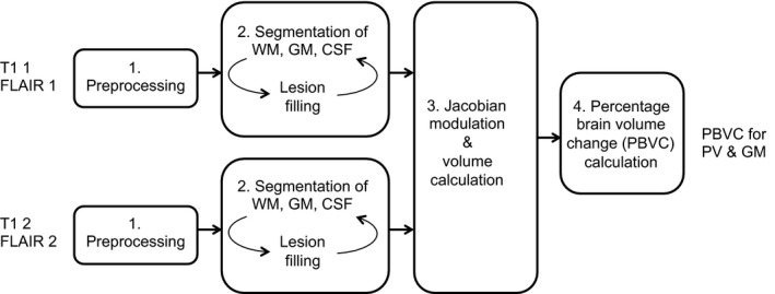
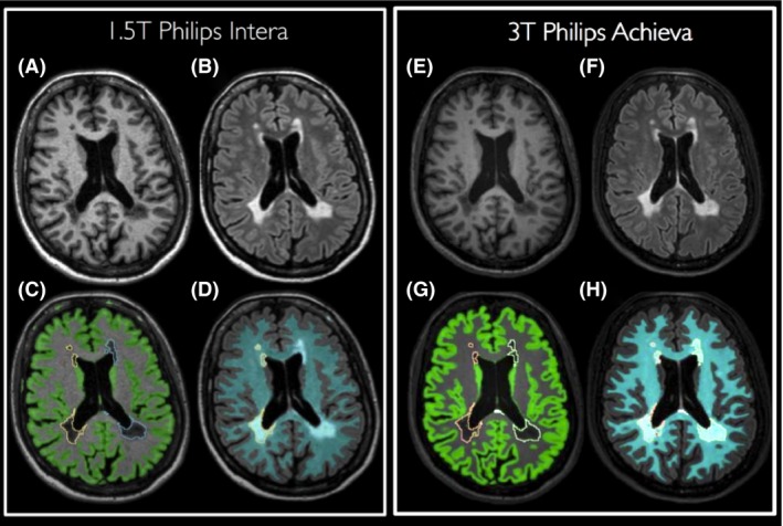
Therefore, in this study 18 MS patients were scanned on the same day on a 1.5T and a 3T scanner. For each scanner, a 3D T1 and a 3D FLAIR were acquired. As no atrophy is expected within 1 day, these datasets can be used to evaluate the median percentage error of the brain volume measurement for gray matter (GM) volume and parenchymal volume (PV) between 1.5T and 3T scanners. The results are obtained with MSmetrix, which is developed especially for use in the MS clinical care path, and compared to Siena (FSL), a widely used software for research purposes.

RESULTS

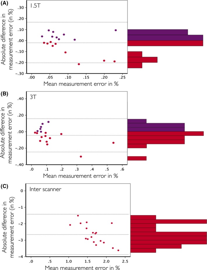
The MSmetrix median percentage error of the brain volume measurement between a 1.5T and a 3T scanner is 0.52% for GM and 0.35% for PV. For Siena this error equals 2.99%. When data of the same scanner are compared, the error is in the order of 0.06-0.08% for both MSmetrix and Siena.

CONCLUSIONS

MSmetrix appears robust on both the 1.5T and 3T systems and the measurement error becomes an order of magnitude higher between scanners with different field strength.

摘要

引言

越来越多的证据表明,脑萎缩是多发性硬化症(MS)病理生理学的一部分,并且与该疾病的多种临床结果相关,包括身体和认知方面。因此,脑萎缩正成为患者随访中的一个重要参数。由于在临床实践中,1.5特斯拉(T)和3T磁共振成像(MRI)系统都用于MS患者的随访,因此出现了关于兼容性以及是否可能需要标准化的问题。

方法

因此,在本研究中,18名MS患者在同一天分别在1.5T和3T扫描仪上进行扫描。对于每台扫描仪,采集了三维T1加权成像和三维液体衰减反转恢复序列(FLAIR)图像。由于预计一天内不会出现萎缩,这些数据集可用于评估1.5T和3T扫描仪之间灰质(GM)体积和实质体积(PV)的脑体积测量的中位百分比误差。结果是使用专门为MS临床护理路径开发的MSmetrix获得的,并与广泛用于研究目的的Siena(FSL)软件进行比较。

结果

1.5T和3T扫描仪之间脑体积测量的MSmetrix中位百分比误差,GM为0.52%,PV为0.35%。对于Siena,该误差为2.99%。当比较同一台扫描仪的数据时,MSmetrix和Siena的误差均在0.06 - 0.08%左右。

结论

MSmetrix在1.5T和3T系统上似乎都很稳定,并且不同场强扫描仪之间的测量误差会高出一个数量级。

https://cdn.ncbi.nlm.nih.gov/pmc/blobs/cc11/4834931/a30160b19806/BRB3-6-e00422-g004.jpg
https://cdn.ncbi.nlm.nih.gov/pmc/blobs/cc11/4834931/259e72b0f177/BRB3-6-e00422-g001.jpg
https://cdn.ncbi.nlm.nih.gov/pmc/blobs/cc11/4834931/8c650d7fa735/BRB3-6-e00422-g002.jpg
https://cdn.ncbi.nlm.nih.gov/pmc/blobs/cc11/4834931/5a90115c52bc/BRB3-6-e00422-g003.jpg
https://cdn.ncbi.nlm.nih.gov/pmc/blobs/cc11/4834931/a30160b19806/BRB3-6-e00422-g004.jpg

相似文献

1
Quantifying brain volumes for Multiple Sclerosis patients follow-up in clinical practice - comparison of 1.5 and 3 Tesla magnetic resonance imaging.在临床实践中对多发性硬化症患者进行随访时量化脑容量——1.5特斯拉和3特斯拉磁共振成像的比较
Brain Behav. 2016 Jan 12;6(2):e00422. doi: 10.1002/brb3.422. eCollection 2016 Feb.
2
Agreement of MSmetrix with established methods for measuring cross-sectional and longitudinal brain atrophy.MSmetrix 与用于测量脑横断面积和纵向萎缩的既定方法的一致性。
Neuroimage Clin. 2017 Jun 30;15:843-853. doi: 10.1016/j.nicl.2017.06.034. eCollection 2017.
3
Measurement of Whole-Brain and Gray Matter Atrophy in Multiple Sclerosis: Assessment with MR Imaging.磁共振成像评估多发性硬化症全脑和灰质萎缩的测量。
Radiology. 2018 Aug;288(2):554-564. doi: 10.1148/radiol.2018172468. Epub 2018 May 1.
4
Whole Brain Volume Measured from 1.5T versus 3T MRI in Healthy Subjects and Patients with Multiple Sclerosis.在健康受试者和多发性硬化症患者中,通过1.5T与3T磁共振成像测量全脑体积。
J Neuroimaging. 2016 Jan-Feb;26(1):62-7. doi: 10.1111/jon.12271. Epub 2015 Jun 28.
5
Feasibility of Brain Atrophy Measurement in Clinical Routine without Prior Standardization of the MRI Protocol: Results from MS-MRIUS, a Longitudinal Observational, Multicenter Real-World Outcome Study in Patients with Relapsing-Remitting MS.临床常规中不进行 MRI 方案标准化而进行脑萎缩测量的可行性:来自 MS-MRIUS 的结果,这是一项在复发缓解型多发性硬化症患者中进行的纵向观察性、多中心真实世界结局研究。
AJNR Am J Neuroradiol. 2018 Feb;39(2):289-295. doi: 10.3174/ajnr.A5442. Epub 2017 Nov 23.
6
Intra- and interscanner variability of magnetic resonance imaging based volumetry in multiple sclerosis.基于磁共振成像的多发性硬化症容积测量的扫描仪内和扫描仪间变异性。
Neuroimage. 2016 Nov 15;142:188-197. doi: 10.1016/j.neuroimage.2016.07.035. Epub 2016 Jul 16.
7
FLAIR-only joint volumetric analysis of brain lesions and atrophy in clinically isolated syndrome (CIS) suggestive of multiple sclerosis.FLAIR 仅用于联合评估疑似多发性硬化症的临床孤立综合征 (CIS) 患者的脑损伤和萎缩的容积分析。
Neuroimage Clin. 2021;29:102542. doi: 10.1016/j.nicl.2020.102542. Epub 2020 Dec 25.
8
Neurological software tool for reliable atrophy measurement (NeuroSTREAM) of the lateral ventricles on clinical-quality T2-FLAIR MRI scans in multiple sclerosis.用于在多发性硬化症的临床质量 T2-FLAIR MRI 扫描上进行可靠的侧脑室萎缩测量的神经学软件工具 (NeuroSTREAM)。
Neuroimage Clin. 2017 Jun 19;15:769-779. doi: 10.1016/j.nicl.2017.06.022. eCollection 2017.
9
Intercontinental validation of brain volume measurements using MSmetrix.使用MSmetrix进行脑容量测量的洲际验证。
Neuroradiol J. 2018 Apr;31(2):147-149. doi: 10.1177/1971400917721521. Epub 2017 Jul 21.
10
Repeatability and reproducibility of FreeSurfer, FSL-SIENAX and SPM brain volumetric measurements and the effect of lesion filling in multiple sclerosis.FreeSurfer、FSL-SIENAX 和 SPM 脑容量测量的可重复性和再现性,以及多发性硬化症中病变填充的影响。
Eur Radiol. 2019 Mar;29(3):1355-1364. doi: 10.1007/s00330-018-5710-x. Epub 2018 Sep 21.

引用本文的文献

1
Uncovering heterogeneous cognitive trajectories in relapsing-remitting multiple sclerosis: a longitudinal study.揭示复发缓解型多发性硬化症中的异质性认知轨迹:一项纵向研究。
Acta Neurol Belg. 2025 Apr;125(2):519-530. doi: 10.1007/s13760-025-02754-7. Epub 2025 Mar 15.
2
Evaluating brain volume segmentation accuracy and reliability of FreeSurfer and Neurophet AQUA at variations in MRI magnetic field strengths.评估 FreeSurfer 和 Neurophet AQUA 在 MRI 磁场强度变化时的脑容量分割准确性和可靠性。
Sci Rep. 2024 Oct 18;14(1):24513. doi: 10.1038/s41598-024-74622-y.
3
Patient-reported outcome measurements in a selective cohort of relapsing-remitting multiple sclerosis patients: relationships with physical disability, cognitive impairment, and MRI-derived metrics.

本文引用的文献

1
Is it time to target no evident disease activity (NEDA) in multiple sclerosis?是时候将目标设定为多发性硬化症的无明显疾病活动(NEDA)了吗?
Mult Scler Relat Disord. 2015 Jul;4(4):329-33. doi: 10.1016/j.msard.2015.04.006. Epub 2015 May 8.
2
Automatic segmentation and volumetry of multiple sclerosis brain lesions from MR images.基于磁共振图像的多发性硬化症脑损伤自动分割与容积测量
Neuroimage Clin. 2015 May 16;8:367-75. doi: 10.1016/j.nicl.2015.05.003. eCollection 2015.
3
Correlation between brain volume change and T2 relaxation time induced by dehydration and rehydration: implications for monitoring atrophy in clinical studies.
在一个复发缓解型多发性硬化症患者的选择性队列中,患者报告的结局测量指标与身体残疾、认知障碍和 MRI 衍生指标的关系。
Acta Neurol Belg. 2023 Jun;123(3):1049-1059. doi: 10.1007/s13760-023-02225-x. Epub 2023 Mar 6.
4
Commercial volumetric MRI reporting tools in multiple sclerosis: a systematic review of the evidence.商业容积磁共振成像报告工具在多发性硬化症中的应用:系统评价证据。
Neuroradiology. 2023 Jan;65(1):5-24. doi: 10.1007/s00234-022-03074-w. Epub 2022 Nov 4.
5
Gadolinium-based contrast agent exposures and physical and cognitive disability in multiple sclerosis.钆基造影剂暴露与多发性硬化症的身体和认知残疾。
J Neuroimaging. 2023 Jan;33(1):85-93. doi: 10.1111/jon.13057. Epub 2022 Oct 1.
6
Brain age as a surrogate marker for cognitive performance in multiple sclerosis.脑龄作为多发性硬化症认知表现的替代标志物。
Eur J Neurol. 2022 Oct;29(10):3039-3049. doi: 10.1111/ene.15473. Epub 2022 Jul 11.
7
Morphometric evaluation of traumatic axonal injury and the correlation with post-traumatic cerebral atrophy and functional outcome.创伤性轴索损伤的形态计量学评估及其与创伤后脑萎缩和功能预后的相关性。
Neuroradiol J. 2022 Aug;35(4):468-476. doi: 10.1177/19714009211049714. Epub 2021 Oct 13.
8
A Novel Digital Care Management Platform to Monitor Clinical and Subclinical Disease Activity in Multiple Sclerosis.一种用于监测多发性硬化症临床和亚临床疾病活动的新型数字护理管理平台。
Brain Sci. 2021 Sep 3;11(9):1171. doi: 10.3390/brainsci11091171.
9
Enlarged perivascular spaces in multiple sclerosis on magnetic resonance imaging: a systematic review and meta-analysis.磁共振成像多发性硬化症中的血管周围间隙扩大:系统评价和荟萃分析。
J Neurol. 2020 Nov;267(11):3199-3212. doi: 10.1007/s00415-020-09971-5. Epub 2020 Jun 13.
10
Diagnosis and management of multiple sclerosis: MRI in clinical practice.多发性硬化的诊断与管理:临床实践中的磁共振成像
J Neurol. 2020 Oct;267(10):2917-2925. doi: 10.1007/s00415-020-09930-0. Epub 2020 May 29.
脱水和再水化引起的脑容量变化与T2弛豫时间之间的相关性:对临床研究中监测萎缩的意义。
Neuroimage Clin. 2014 Aug 23;6:166-70. doi: 10.1016/j.nicl.2014.08.014. eCollection 2014.
4
The SIENA/FSL whole brain atrophy algorithm is no more reproducible at 3T than 1.5 T for Alzheimer's disease.SIENA/FSL 全脑萎缩算法在 3T 与 1.5T 场强对于阿尔茨海默病均不可重现。
Psychiatry Res. 2014 Oct 30;224(1):14-21. doi: 10.1016/j.pscychresns.2014.07.002. Epub 2014 Jul 14.
5
Accurate GM atrophy quantification in MS using lesion-filling with co-registered 2D lesion masks.在多发性硬化症中,使用共配准的二维病变掩码进行病变填充来准确量化脑灰质萎缩。
Neuroimage Clin. 2014 Jan 18;4:366-73. doi: 10.1016/j.nicl.2014.01.004. eCollection 2014.
6
Pathology of multiple sclerosis and related inflammatory demyelinating diseases.多发性硬化症及相关炎性脱髓鞘疾病的病理学
Handb Clin Neurol. 2014;122:15-58. doi: 10.1016/B978-0-444-52001-2.00002-9.
7
Brain atrophy and lesion load predict long term disability in multiple sclerosis.脑萎缩和病灶负荷可预测多发性硬化的长期残疾。
J Neurol Neurosurg Psychiatry. 2013 Oct;84(10):1082-91. doi: 10.1136/jnnp-2012-304094. Epub 2013 Mar 23.
8
Optimizing parameter choice for FSL-Brain Extraction Tool (BET) on 3D T1 images in multiple sclerosis.优化多发性硬化症 3D T1 图像 FSL-Brain Extraction Tool(BET)的参数选择。
Neuroimage. 2012 Jul 16;61(4):1484-94. doi: 10.1016/j.neuroimage.2012.03.074. Epub 2012 Mar 30.
9
Association between pathological and MRI findings in multiple sclerosis.多发性硬化的病理与 MRI 表现的相关性。
Lancet Neurol. 2012 Apr;11(4):349-60. doi: 10.1016/S1474-4422(12)70003-0. Epub 2012 Mar 19.
10
Evaluating and reducing the impact of white matter lesions on brain volume measurements.评估和降低脑白质病变对脑容量测量的影响。
Hum Brain Mapp. 2012 Sep;33(9):2062-71. doi: 10.1002/hbm.21344. Epub 2011 Aug 31.