Simsekyilmaz Sakine, Liehn Elisa A, Weinandy Stefan, Schreiber Fabian, Megens Remco T A, Theelen Wendy, Smeets Ralf, Jockenhövel Stefan, Gries Thomas, Möller Martin, Klee Doris, Weber Christian, Zernecke Alma
Institute for Molecular Cardiovascular Research, University Hospital Aachen, RWTH Aachen University, Aachen, Germany.
Institute of Technical and Macromolecular Chemistry, RWTH Aachen University, Aachen, Germany.
PLoS One. 2016 May 18;11(5):e0155829. doi: 10.1371/journal.pone.0155829. eCollection 2016.
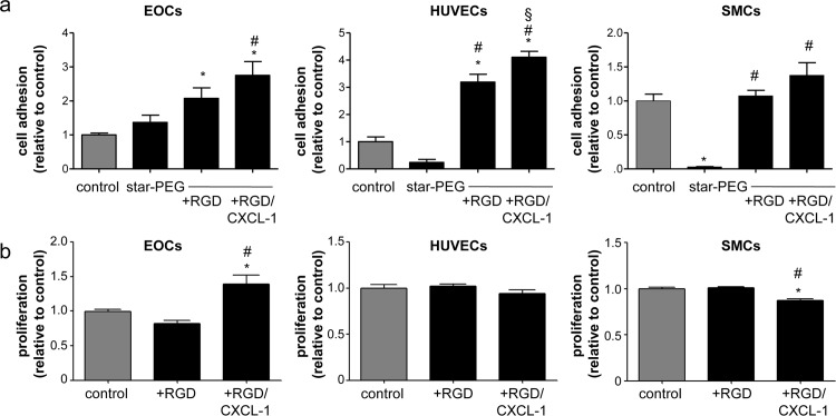
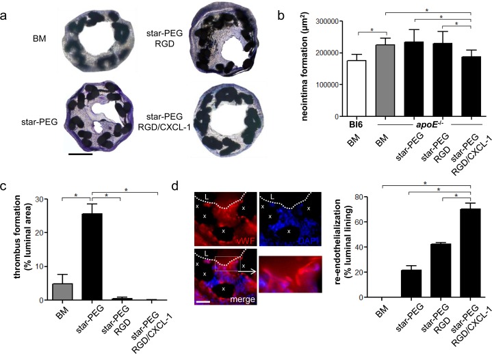
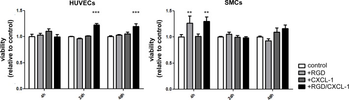
Atherosclerotic lesions that critically narrow the artery can necessitate an angioplasty and stent implantation. Long-term therapeutic effects, however, are limited by excessive arterial remodeling. We here employed a miniaturized nitinol-stent coated with star-shaped polyethylenglycole (star-PEG), and evaluated its bio-functionalization with RGD and CXCL1 for improving in-stent stenosis after implantation into carotid arteries of mice. Nitinol foils or stents (bare metal) were coated with star-PEG, and bio-functionalized with RGD, or RGD/CXCL1. Cell adhesion to star-PEG-coated nitinol foils was unaltered or reduced, whereas bio-functionalization with RGD but foremost RGD/CXCL1 increased adhesion of early angiogenic outgrowth cells (EOCs) and endothelial cells but not smooth muscle cells when compared with bare metal foils. Stimulation of cells with RGD/CXCL1 furthermore increased the proliferation of EOCs. In vivo, bio-functionalization with RGD/CXCL1 significantly reduced neointima formation and thrombus formation, and increased re-endothelialization in apoE-/- carotid arteries compared with bare-metal nitinol stents, star-PEG-coated stents, and stents bio-functionalized with RGD only. Bio-functionalization of star-PEG-coated nitinol-stents with RGD/CXCL1 reduced in-stent neointima formation. By supporting the adhesion and proliferation of endothelial progenitor cells, RGD/CXCL1 coating of stents may help to accelerate endothelial repair after stent implantation, and thus may harbor the potential to limit the complication of in-stent restenosis in clinical approaches.
严重使动脉狭窄的动脉粥样硬化病变可能需要进行血管成形术和支架植入。然而,长期治疗效果受到过度动脉重塑的限制。我们在此采用了一种涂有星形聚乙二醇(star-PEG)的小型镍钛诺支架,并评估了其用RGD和CXCL1进行生物功能化处理后对植入小鼠颈动脉后支架内狭窄的改善情况。镍钛诺箔片或支架(裸金属)用star-PEG进行涂层,并分别用RGD或RGD/CXCL1进行生物功能化处理。与裸金属箔片相比,细胞对涂有star-PEG的镍钛诺箔片的黏附未改变或减少,而用RGD尤其是RGD/CXCL1进行生物功能化处理则增加了早期血管生成外植细胞(EOCs)和内皮细胞的黏附,但对平滑肌细胞无此作用。用RGD/CXCL1刺激细胞还能增加EOCs的增殖。在体内,与裸金属镍钛诺支架、涂有star-PEG的支架以及仅用RGD进行生物功能化处理的支架相比,用RGD/CXCL1进行生物功能化处理显著减少了载脂蛋白E基因敲除小鼠颈动脉内的新生内膜形成和血栓形成,并增加了再内皮化。用RGD/CXCL1对涂有star-PEG的镍钛诺支架进行生物功能化处理可减少支架内新生内膜形成。通过支持内皮祖细胞的黏附和增殖,支架的RGD/CXCL1涂层可能有助于加速支架植入后的内皮修复,因此在临床方法中可能具有限制支架内再狭窄并发症的潜力。