• 文献检索
  • 文档翻译
  • 深度研究
  • 学术资讯
  • Suppr Zotero 插件Zotero 插件
  • 邀请有礼
  • 套餐&价格
  • 历史记录
应用&插件
Suppr Zotero 插件Zotero 插件浏览器插件Mac 客户端Windows 客户端微信小程序
定价
高级版会员购买积分包购买API积分包
服务
文献检索文档翻译深度研究API 文档MCP 服务
关于我们
关于 Suppr公司介绍联系我们用户协议隐私条款
关注我们

Suppr 超能文献

核心技术专利:CN118964589B侵权必究
粤ICP备2023148730 号-1Suppr @ 2026

文献检索

告别复杂PubMed语法,用中文像聊天一样搜索,搜遍4000万医学文献。AI智能推荐,让科研检索更轻松。

立即免费搜索

文件翻译

保留排版,准确专业,支持PDF/Word/PPT等文件格式,支持 12+语言互译。

免费翻译文档

深度研究

AI帮你快速写综述,25分钟生成高质量综述,智能提取关键信息,辅助科研写作。

立即免费体验

技术与青光眼可疑患者

Technology and the Glaucoma Suspect.

作者信息

Blumberg Dana M, De Moraes Carlos Gustavo, Liebmann Jeffrey M, Garg Reena, Chen Cynthia, Theventhiran Alex, Hood Donald C

机构信息

Bernard and Shirlee Brown Glaucoma Research Laboratory, Department of Ophthalmology, Edward S. Harkness Eye Institute, Columbia University Medical Center, New York, New York, United States.

Department of Ophthalmology, New York Eye and Ear Infirmary of Mount Sinai Department of Ophthalmology, New York, New York, United States.

出版信息

Invest Ophthalmol Vis Sci. 2016 Jul 1;57(9):OCT80-5. doi: 10.1167/iovs.15-18931.

DOI:10.1167/iovs.15-18931
PMID:27409509
原文链接:https://pmc.ncbi.nlm.nih.gov/articles/PMC5995486/
Abstract

PURPOSE

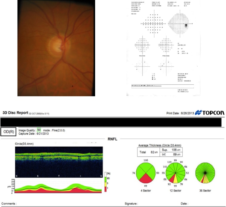
To determine and compare the diagnostic performance of spectral-domain optical coherence tomography (SD-OCT), stereoscopic disc photographs, and automated perimetry as assessed by a group of glaucoma specialists in differentiating individuals with early glaucoma from suspects.

METHODS

Forty-six eyes (46 patients) with suspicious optic nerves had previously undergone SD-OCT scans, 24-2 visual fields (VFs), and optic disc photographs. The average VF mean deviation was -1.97 ± 2.09 (SD) dB. Four glaucoma specialists examined the 138 individual diagnostic tests and classified the patient as likely glaucomatous or nonglaucomatous based on the results of a single test. The diagnostic performances of each of the three tests were compared to a previously determined reference standard, based on the consensus of a separate panel of four glaucoma specialists who examined all three tests together.

RESULTS

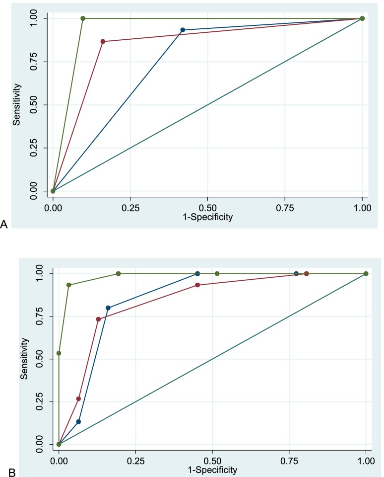
Among the four specialists, the interobserver agreement across the three diagnostic tests was poor for VF and photos, with kappa (κ) values of 0.13 and 0.16, respectively, and moderate for OCT, with κ value of 0.40. Using panel consensus as reference standard, OCT had the highest discriminative ability, with an area under the curve (AUC) of 0.99 (95% 0.96-1.0) compared to photograph AUC 0.85 (95% 0.73-0.96) and VF AUC 0.86 (95% 0.76-0.96), suggestive of closer performance to that of a group of glaucoma specialists.

CONCLUSIONS

Compared to VF and disc photography, SD-OCT, when used alone, had better internal agreement as well as better agreement with the consensus of clinicians using all available data. Future studies should evaluate best practices for SD-OCT interpretation.

摘要

目的

由一组青光眼专家评估,以确定并比较频域光学相干断层扫描(SD - OCT)、立体视盘照片和自动视野计在鉴别早期青光眼患者与可疑青光眼患者时的诊断性能。

方法

46只患有可疑视神经病变的眼睛(46例患者)此前已接受SD - OCT扫描、24 - 2视野(VF)检查和视盘照片拍摄。平均VF平均偏差为-1.97±2.09(标准差)dB。四位青光眼专家检查了这138项个体诊断测试,并根据单一测试结果将患者分类为可能患有青光眼或非青光眼。基于由四位共同检查所有三项测试的青光眼专家组成的独立小组的共识,将这三项测试各自的诊断性能与先前确定的参考标准进行比较。

结果

在这四位专家中,对于VF和照片,三项诊断测试的观察者间一致性较差,kappa(κ)值分别为0.13和0.16,而对于OCT,一致性为中等,κ值为0.40。以小组共识作为参考标准,OCT具有最高的鉴别能力,曲线下面积(AUC)为0.99(95% 0.96 - 1.0),相比之下,照片AUC为0.85(95% 0.73 - 0.96),VF AUC为0.86(95% 0.76 - 0.96),这表明其性能更接近一组青光眼专家的表现。

结论

与VF和视盘摄影相比,单独使用SD - OCT时具有更好的内部一致性,并且与使用所有可用数据的临床医生共识的一致性更好。未来的研究应评估SD - OCT解读的最佳实践。

https://cdn.ncbi.nlm.nih.gov/pmc/blobs/d5c0/5995486/7babaf7e96a5/i1552-5783-57-9-OCT80-f02.jpg
https://cdn.ncbi.nlm.nih.gov/pmc/blobs/d5c0/5995486/5ffacc8d8e58/i1552-5783-57-9-OCT80-f01.jpg
https://cdn.ncbi.nlm.nih.gov/pmc/blobs/d5c0/5995486/7babaf7e96a5/i1552-5783-57-9-OCT80-f02.jpg
https://cdn.ncbi.nlm.nih.gov/pmc/blobs/d5c0/5995486/5ffacc8d8e58/i1552-5783-57-9-OCT80-f01.jpg
https://cdn.ncbi.nlm.nih.gov/pmc/blobs/d5c0/5995486/7babaf7e96a5/i1552-5783-57-9-OCT80-f02.jpg

相似文献

1
Technology and the Glaucoma Suspect.技术与青光眼可疑患者
Invest Ophthalmol Vis Sci. 2016 Jul 1;57(9):OCT80-5. doi: 10.1167/iovs.15-18931.
2
Application of optical coherence tomography in glaucoma suspect eyes.光学相干断层扫描在青光眼可疑眼中的应用。
Clin Exp Optom. 2012 Jan;95(1):78-88. doi: 10.1111/j.1444-0938.2011.00654.x. Epub 2011 Oct 10.
3
Agreement among spectral-domain optical coherence tomography, standard automated perimetry, and stereophotography in the detection of glaucoma progression.光谱域光学相干断层扫描、标准自动视野计和立体摄影在青光眼进展检测中的一致性。
Invest Ophthalmol Vis Sci. 2015 Jan 27;56(2):1253-60. doi: 10.1167/iovs.14-14994.
4
Retinal nerve fiber layer imaging with spectral-domain optical coherence tomography a study on diagnostic agreement with Heidelberg Retinal Tomograph.频域光学相干断层扫描视网膜神经纤维层成像-海德堡视网膜断层扫描仪诊断符合度的研究。
Ophthalmology. 2010 Feb;117(2):267-74. doi: 10.1016/j.ophtha.2009.06.061. Epub 2009 Dec 6.
5
Validation of the structure-function correlation report from the heidelberg edge perimeter and spectral-domain optical coherence tomography.海德堡周边视野计与频域光学相干断层扫描结构-功能相关性报告的验证
Int Ophthalmol. 2019 Mar;39(3):533-540. doi: 10.1007/s10792-018-0836-z. Epub 2018 Feb 2.
6
Structure-Function Relationship between Flicker-Defined Form Perimetry and Spectral-Domain Optical Coherence Tomography in Glaucoma Suspects.青光眼可疑患者中闪烁定义视野检查与频域光学相干断层扫描之间的结构-功能关系
Curr Eye Res. 2017 Mar;42(3):418-423. doi: 10.1080/02713683.2016.1190848. Epub 2016 Jul 15.
7
The Relative Odds of Progressing by Structural and Functional Tests in Glaucoma.青光眼患者通过结构和功能检查进展的相对几率
Invest Ophthalmol Vis Sci. 2016 Jul 1;57(9):OCT421-8. doi: 10.1167/iovs.15-18940.
8
Progression of retinal nerve fiber layer thinning in glaucoma assessed by cirrus optical coherence tomography-guided progression analysis.基于 Cirrus 光学相干断层扫描引导的进展分析评估青光眼的视网膜神经纤维层变薄进展。
Curr Eye Res. 2013 Mar;38(3):386-95. doi: 10.3109/02713683.2012.742913.
9
Correlation of frequency-doubling perimetry with retinal nerve fiber layer thickness and optic disc size in ocular hypertensives and glaucoma suspects.在高眼压症和青光眼疑似患者中,频域光相干断层扫描与视网膜神经纤维层厚度和视盘大小的相关性。
J Glaucoma. 2011 Aug;20(6):366-70. doi: 10.1097/IJG.0b013e3181eda96e.
10
Detecting early glaucoma by assessment of retinal nerve fiber layer thickness and visual function.通过评估视网膜神经纤维层厚度和视觉功能来检测早期青光眼。
Invest Ophthalmol Vis Sci. 2001 Aug;42(9):1993-2003.

引用本文的文献

1
Evaluating Diagnostic Concordance in Primary Open-Angle Glaucoma Among Academic Glaucoma Subspecialists.评估学术性青光眼亚专科医生对原发性开角型青光眼的诊断一致性。
Diagnostics (Basel). 2024 Nov 3;14(21):2460. doi: 10.3390/diagnostics14212460.
2
Automated Optic Nerve Head Hemoglobin Measurements versus General Ophthalmologists Evaluation to Differentiate Glaucomatous from Large Physiological Cupping: A Diagnostic Performance Comparative Study.自动测量视神经乳头血红蛋白与普通眼科医生评估在鉴别青光眼性大视杯与生理性大视杯方面的比较:一项诊断性能对比研究。
Clin Ophthalmol. 2024 Jul 16;18:2073-2081. doi: 10.2147/OPTH.S466349. eCollection 2024.
3

本文引用的文献

1
Management of advanced glaucoma: Characterization and monitoring.晚期青光眼的管理:特征与监测
Surv Ophthalmol. 2016 Sep-Oct;61(5):597-615. doi: 10.1016/j.survophthal.2016.03.006. Epub 2016 Mar 25.
2
Predicting Development of Glaucomatous Visual Field Conversion Using Baseline Fourier-Domain Optical Coherence Tomography.利用基线傅里叶域光学相干断层扫描预测青光眼视野转换的发展
Am J Ophthalmol. 2016 Mar;163:29-37. doi: 10.1016/j.ajo.2015.11.029. Epub 2015 Nov 26.
3
Evaluation of a One-Page Report to Aid in Detecting Glaucomatous Damage.
Classifying glaucoma exclusively with OCT: comparison of three clustering algorithms derived from machine learning.
仅用 OCT 对青光眼进行分类:三种源于机器学习的聚类算法的比较。
Eye (Lond). 2024 Apr;38(5):841-846. doi: 10.1038/s41433-023-02785-5. Epub 2023 Oct 19.
4
Agreement between Five Experts and the Laguna ONhE Automatic Colourimetric Application Interpreting the Glaucomatous Aspect of the Optic Nerve.五位专家与拉古纳ONhE自动比色应用在解读视神经青光眼特征方面的一致性。
J Clin Med. 2023 Aug 24;12(17):5485. doi: 10.3390/jcm12175485.
5
Amsler Grid Versus 10-2 Test in Primary Open Angle Glaucoma.原发性开角型青光眼患者中阿姆斯勒方格表与10-2视野检查的比较
J West Afr Coll Surg. 2023 Apr-Jun;13(2):92-99. doi: 10.4103/jwas.jwas_275_22. Epub 2023 Mar 20.
6
A Neural Network for Automated Image Quality Assessment of Optic Disc Photographs.用于视盘照片自动图像质量评估的神经网络。
J Clin Med. 2023 Feb 3;12(3):1217. doi: 10.3390/jcm12031217.
7
Prediction of visual field defects from macular optical coherence tomography in glaucoma using cluster analysis.利用聚类分析从青光眼的黄斑光学相干断层扫描中预测视野缺损。
Ophthalmic Physiol Opt. 2022 Sep;42(5):948-964. doi: 10.1111/opo.12997. Epub 2022 May 22.
8
Accuracy of Using Generative Adversarial Networks for Glaucoma Detection: Systematic Review and Bibliometric Analysis.使用生成对抗网络进行青光眼检测的准确性:系统评价和文献计量分析。
J Med Internet Res. 2021 Sep 21;23(9):e27414. doi: 10.2196/27414.
9
[Glaucoma care in Germany-Results of a survey among German ophthalmologists-Part 1: diagnostics].[德国的青光眼护理——德国眼科医生的一项调查结果——第1部分:诊断]
Ophthalmologe. 2022 Jan;119(1):38-45. doi: 10.1007/s00347-021-01352-1. Epub 2021 Mar 17.
10
Comparison of spectral domain optical coherence tomography parameters between disc suspects and "pre-perimetric" glaucomatous discs classified on disc photo.比较基于眼底照相的可疑盘与“超视野”青光眼盘的光谱域光学相干断层扫描参数
Indian J Ophthalmol. 2021 Mar;69(3):603-610. doi: 10.4103/ijo.IJO_1309_20.
一份用于辅助检测青光眼性损伤的单页报告的评估
Transl Vis Sci Technol. 2014 Dec 17;3(6):8. doi: 10.1167/tvst.3.6.8. eCollection 2014 Oct.
4
Additive diagnostic role of imaging in glaucoma: optical coherence tomography and retinal nerve fiber layer photography.青光眼成像的附加诊断作用:光学相干断层扫描和视网膜神经纤维层摄影
Invest Ophthalmol Vis Sci. 2014 Nov 20;55(12):8024-30. doi: 10.1167/iovs.14-15237.
5
Visual fields and OCT role in diagnosis of glaucoma.视野检查和光学相干断层扫描在青光眼诊断中的作用。
Optom Vis Sci. 2014 Nov;91(11):1312-9. doi: 10.1097/OPX.0000000000000394.
6
On improving the use of OCT imaging for detecting glaucomatous damage.提高 OCT 成像检测青光眼损伤的应用。
Br J Ophthalmol. 2014 Jul;98 Suppl 2(Suppl 2):ii1-9. doi: 10.1136/bjophthalmol-2014-305156.
7
Comparison of different spectral domain OCT scanning protocols for diagnosing preperimetric glaucoma.不同谱域 OCT 扫描方案诊断青光眼前期的比较。
Invest Ophthalmol Vis Sci. 2013 May 13;54(5):3417-25. doi: 10.1167/iovs.13-11676.
8
Glaucomatous damage of the macula.黄斑青光眼损害。
Prog Retin Eye Res. 2013 Jan;32:1-21. doi: 10.1016/j.preteyeres.2012.08.003. Epub 2012 Sep 17.
9
Prediction of functional loss in glaucoma from progressive optic disc damage.从进行性视盘损害预测青光眼的功能丧失
Arch Ophthalmol. 2009 Oct;127(10):1250-6. doi: 10.1001/archophthalmol.2009.276.
10
Comparison of quantitative imaging devices and subjective optic nerve head assessment by general ophthalmologists to differentiate normal from glaucomatous eyes.定量成像设备与普通眼科医生的主观视神经乳头评估用于鉴别正常眼与青光眼性眼的比较。
J Glaucoma. 2009 Mar;18(3):253-61. doi: 10.1097/IJG.0b013e31818153da.