Mayer Britta, Soppert Josefin, Kraemer Sandra, Schemmel Sabrina, Beckers Christian, Bleilevens Christian, Rossaint Rolf, Coburn Mark, Goetzenich Andreas, Stoppe Christian
Department of Thoracic & Cardiovascular Surgery, University Hospital RWTH, 52074 Aachen, Germany.
Department of Anesthesiology, University Hospital RWTH, 52074 Aachen, Germany.
Int J Mol Sci. 2016 Jul 19;17(7):1159. doi: 10.3390/ijms17071159.
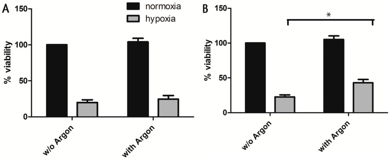
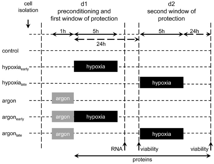
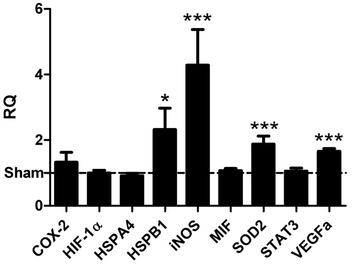
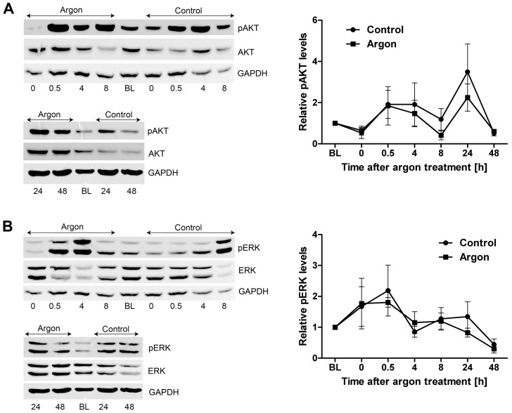
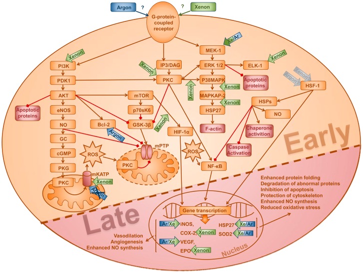
Increasing evidence indicates that argon has organoprotective properties. So far, the underlying mechanisms remain poorly understood. Therefore, we investigated the effect of argon preconditioning in cardiomyocytes within the first and second window of preconditioning. Primary isolated cardiomyocytes from neonatal rats were subjected to 50% argon for 1 h, and subsequently exposed to a sublethal dosage of hypoxia (<1% O₂) for 5 h either within the first (0-3 h) or second window (24-48 h) of preconditioning. Subsequently, the cell viability and proliferation was measured. The argon-induced effects were assessed by evaluation of mRNA and protein expression after preconditioning. Argon preconditioning did not show any cardioprotective effects in the early window of preconditioning, whereas it leads to a significant increase of cell viability 24 h after preconditioning compared to untreated cells (p = 0.015) independent of proliferation. Argon-preconditioning significantly increased the mRNA expression of heat shock protein (HSP) B1 (HSP27) (p = 0.048), superoxide dismutase 2 (SOD2) (p = 0.001), vascular endothelial growth factor (VEGF) (p < 0.001) and inducible nitric oxide synthase (iNOS) (p = 0.001). No difference was found with respect to activation of pro-survival kinases in the early and late window of preconditioning. The findings provide the first evidence of argon-induced effects on the survival of cardiomyocytes during the second window of preconditioning, which may be mediated through the induction of HSP27, SOD2, VEGF and iNOS.
越来越多的证据表明氩气具有器官保护特性。到目前为止,其潜在机制仍知之甚少。因此,我们研究了在预处理的第一和第二时间窗内氩气预处理对心肌细胞的影响。将新生大鼠原代分离的心肌细胞置于50%氩气环境中1小时,随后在预处理的第一时间窗(0 - 3小时)或第二时间窗(24 - 48小时)内暴露于亚致死剂量的低氧环境(<1% O₂)中5小时。随后,测量细胞活力和增殖情况。通过评估预处理后mRNA和蛋白质表达来评估氩气诱导的效应。氩气预处理在预处理的早期时间窗未显示出任何心脏保护作用,而与未处理的细胞相比,预处理24小时后其导致细胞活力显著增加(p = 0.015),且与增殖无关。氩气预处理显著增加了热休克蛋白(HSP)B1(HSP27)(p = 0.048)、超氧化物歧化酶2(SOD2)(p = 0.001)、血管内皮生长因子(VEGF)(p < 0.001)和诱导型一氧化氮合酶(iNOS)(p = 0.001)的mRNA表达。在预处理的早期和晚期时间窗内,促存活激酶的激活方面未发现差异。这些发现首次证明了在预处理的第二时间窗内氩气对心肌细胞存活有诱导作用,这可能是通过诱导HSP27、SOD2、VEGF和iNOS介导的。