Physical Sciences of Imaging for Biomedical Sciences (PSIBS) Doctoral Training Centre, College of Engineering &Physical Sciences, University of Birmingham, Birmingham, B15 2TT, UK.
School of Dentistry, Institute of Clinical Sciences, College of Medical and Dental Sciences, University of Birmingham, Mill Pool Way, Birmingham, B5 7EG, UK.
Sci Rep. 2016 Sep 7;6:32694. doi: 10.1038/srep32694.
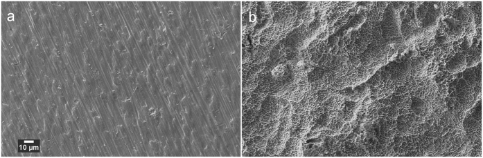
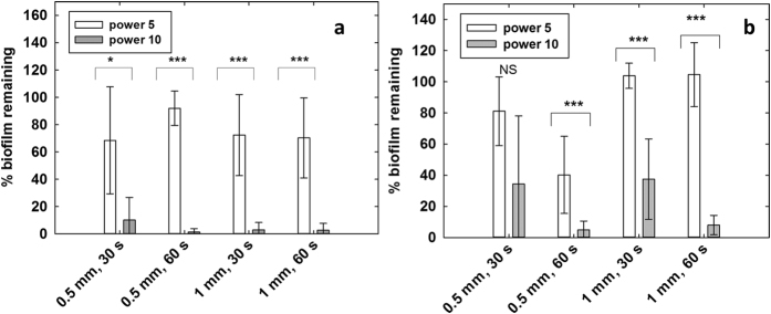
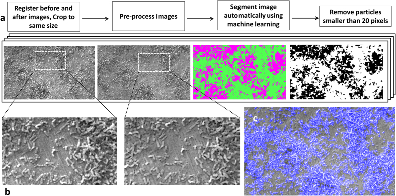
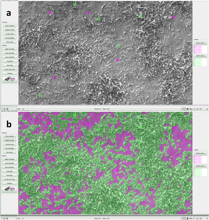
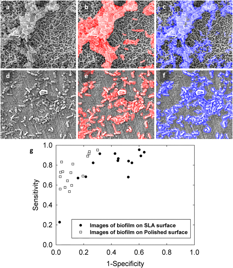
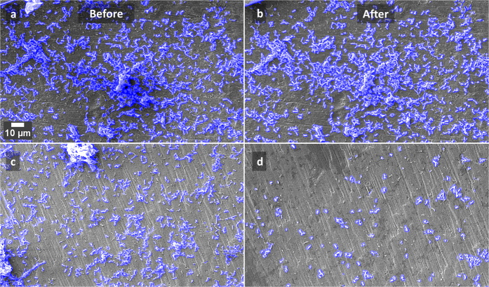
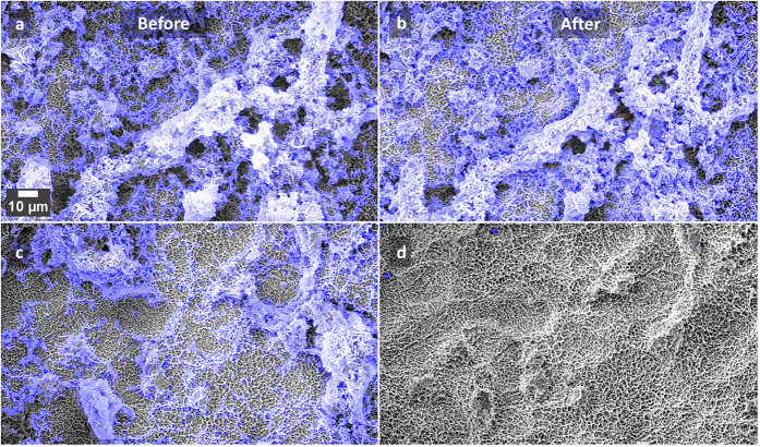
Biofilm accumulation on biomaterial surfaces is a major health concern and significant research efforts are directed towards producing biofilm resistant surfaces and developing biofilm removal techniques. To accurately evaluate biofilm growth and disruption on surfaces, accurate methods which give quantitative information on biofilm area are needed, as current methods are indirect and inaccurate. We demonstrate the use of machine learning algorithms to segment biofilm from scanning electron microscopy images. A case study showing disruption of biofilm from rough dental implant surfaces using cavitation bubbles from an ultrasonic scaler is used to validate the imaging and analysis protocol developed. Streptococcus mutans biofilm was disrupted from sandblasted, acid etched (SLA) Ti discs and polished Ti discs. Significant biofilm removal occurred due to cavitation from ultrasonic scaling (p < 0.001). The mean sensitivity and specificity values for segmentation of the SLA surface images were 0.80 ± 0.18 and 0.62 ± 0.20 respectively and 0.74 ± 0.13 and 0.86 ± 0.09 respectively for polished surfaces. Cavitation has potential to be used as a novel way to clean dental implants. This imaging and analysis method will be of value to other researchers and manufacturers wishing to study biofilm growth and removal.
生物膜在生物材料表面的积累是一个主要的健康问题,因此人们投入了大量的研究努力来生产抗生物膜表面并开发去除生物膜的技术。为了准确评估表面上生物膜的生长和破坏,需要使用能够提供生物膜面积定量信息的准确方法,因为目前的方法是间接的且不准确的。我们展示了使用机器学习算法从扫描电子显微镜图像中分割生物膜的方法。使用超声洁牙器中的空化气泡从粗糙的牙科植入物表面破坏生物膜的案例研究用于验证所开发的成像和分析方案。变形链球菌生物膜从喷砂酸蚀 (SLA) Ti 盘和抛光 Ti 盘上脱落。由于超声洁牙产生的空化作用,生物膜的去除效果显著(p<0.001)。SLA 表面图像分割的平均灵敏度和特异性值分别为 0.80±0.18 和 0.62±0.20,抛光表面的相应值分别为 0.74±0.13 和 0.86±0.09。空化具有作为一种清洁牙科植入物的新方法的潜力。这种成像和分析方法将对其他希望研究生物膜生长和去除的研究人员和制造商具有价值。