Straub Christoph, Saulnier Jessica Lizette, Bègue Aurelien, Feng Danielle D, Huang Kee Wui, Sabatini Bernardo Luis
Howard Hughes Medical Institute, Department of Neurobiology, Harvard Medical School, 220 Longwood Ave., Boston, MA 02115, USA.
Howard Hughes Medical Institute, Department of Neurobiology, Harvard Medical School, 220 Longwood Ave., Boston, MA 02115, USA.
Neuron. 2016 Oct 5;92(1):84-92. doi: 10.1016/j.neuron.2016.09.007.
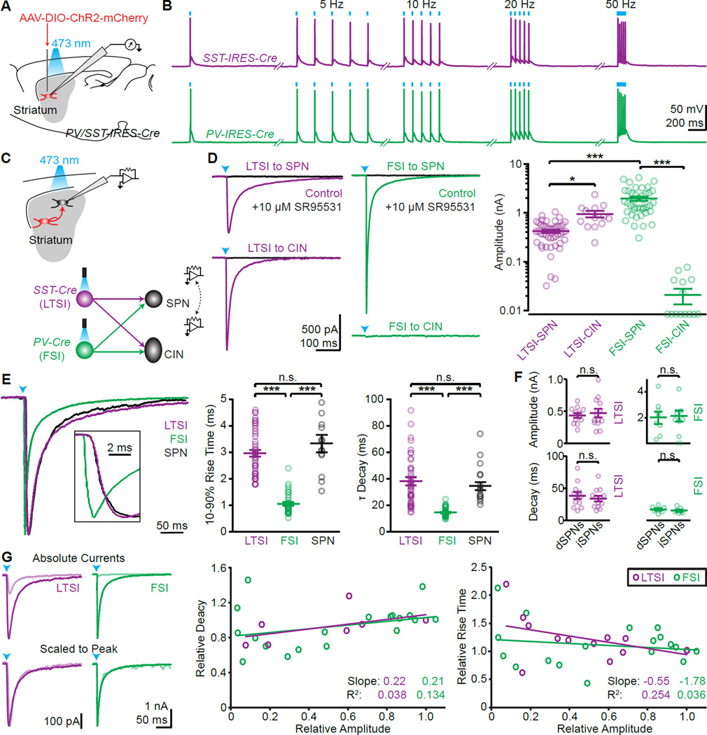
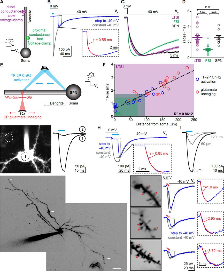
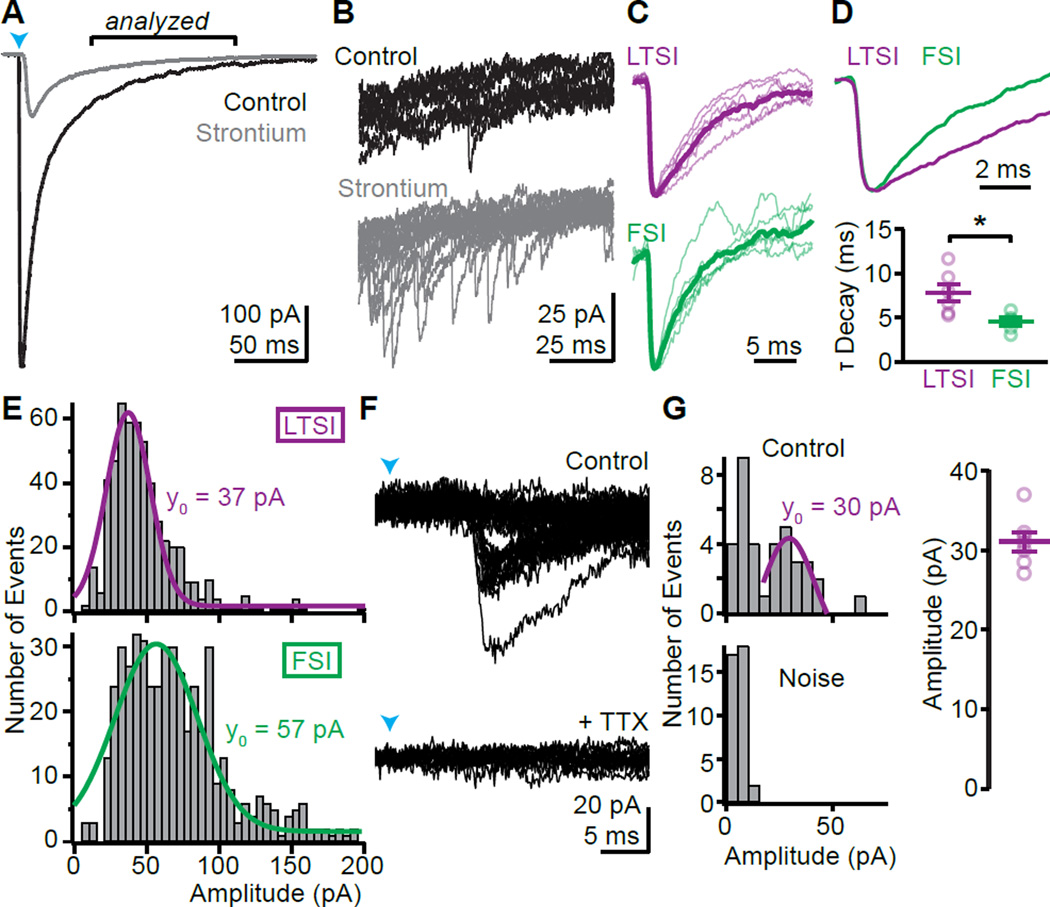
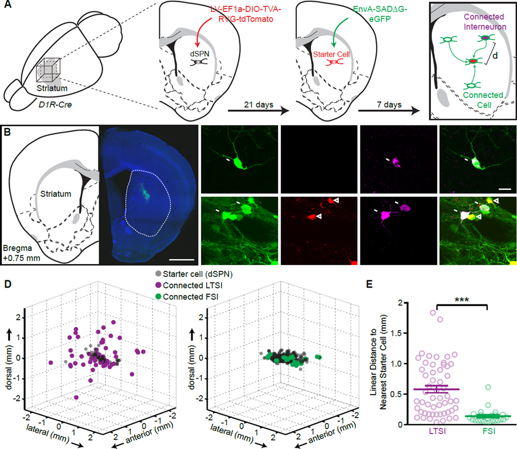
The striatum, the entry nucleus of the basal ganglia, lacks laminar or columnar organization of its principal cells; nevertheless, functional data suggest that it is spatially organized. Here we examine whether the connectivity and synaptic organization of striatal GABAergic interneurons contributes to such spatial organization. Focusing on the two main classes of striatal GABAergic interneurons (fast-spiking interneurons [FSIs] and low-threshold-spiking interneurons [LTSIs]), we apply a combination of optogenetics and viral tracing approaches to dissect striatal microcircuits in mice. Our results reveal fundamental differences between the synaptic organizations of both interneuron types. FSIs target exclusively striatal projection neurons (SPNs) within close proximity and form strong synapses on the proximal somatodendritic region. In contrast, LTSIs target both SPNs and cholinergic interneurons, and synaptic connections onto SPNs are made exclusively over long distances and onto distal dendrites. These results suggest fundamentally different functions of FSIs and LTSIs in shaping striatal output.
纹状体作为基底神经节的输入核,其主要细胞缺乏层状或柱状组织;然而,功能数据表明它在空间上是有组织的。在这里,我们研究纹状体GABA能中间神经元的连接性和突触组织是否有助于这种空间组织。聚焦于纹状体GABA能中间神经元的两类主要细胞(快发放中间神经元[FSIs]和低阈值发放中间神经元[LTSIs]),我们应用光遗传学和病毒示踪方法相结合来剖析小鼠的纹状体微回路。我们的结果揭示了这两种中间神经元类型在突触组织上的根本差异。FSIs仅靶向紧邻的纹状体投射神经元(SPNs),并在近端树突棘区域形成强突触。相比之下,LTSIs既靶向SPNs,也靶向胆碱能中间神经元,并且与SPNs的突触连接仅在远距离且在远端树突上形成。这些结果表明FSIs和LTSIs在塑造纹状体输出方面具有根本不同的功能。