Brunot-Gohin Céline, Duval Jean-Luc, Verbeke Sandra, Belanger Kayla, Pezron Isabelle, Kugel Gérard, Laurent-Maquin Dominique, Gangloff Sophie, Egles Christophe
Laboratory of Biomaterials and Bone Site Inflammation, University of Reims Champagne-Ardenne, Reims, France.; Laboratory of Biomechanics and Bioengineering, Research Center of Royallieu, University of Technology of Compiègne, Sorbonne Universities, Compiègne, France.; University of Reims Champagne-Ardenne, Faculty of Odontology, Reims, France.
Laboratory of Biomechanics and Bioengineering, Research Center of Royallieu, University of Technology of Compiègne, Sorbonne Universities, Compiègne, France .
J Periodontal Implant Sci. 2016 Dec;46(6):362-371. doi: 10.5051/jpis.2016.46.6.362. Epub 2016 Dec 27.
The increasing demand for esthetically pleasing results has contributed to the use of ceramics for dental implant abutments. The aim of this study was to compare the biological response of epithelial tissue cultivated on lithium disilicate (LS) and zirconium oxide (ZrO) ceramics. Understanding the relevant physicochemical and mechanical properties of these ceramics will help identify the optimal material for facilitating gingival wound closure.
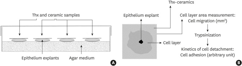
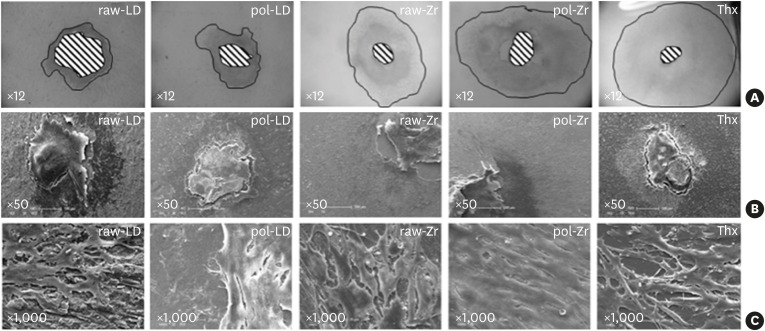
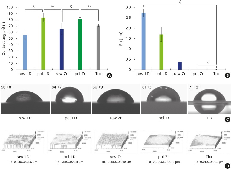
Both biomaterials were prepared with 2 different surface treatments: raw and polished. Their physicochemical characteristics were analyzed by contact angle measurements, scanning white-light interferometry, and scanning electron microscopy. An organotypic culture was then performed using a chicken epithelium model to simulate peri-implant soft tissue. We measured the contact angle, hydrophobicity, and roughness of the materials as well as the tissue behavior at their surfaces (cell migration and cell adhesion).
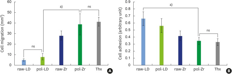
The best cell migration was observed on ZrO ceramic. Cell adhesion was also drastically lower on the polished ZrO ceramic than on both the raw and polished LS. Evaluating various surface topographies of LS showed that increasing surface roughness improved cell adhesion, leading to an increase of up to 13%.
Our results demonstrate that a biomaterial, here LS, can be modified using simple surface changes in order to finely modulate soft tissue adhesion. Strong adhesion at the abutment associated with weak migration assists in gingival wound healing. On the same material, polishing can reduce cell adhesion without drastically modifying cell migration. A comparison of LS and ZrO ceramic showed that LS was more conducive to creating varying tissue reactions. Our results can help dental surgeons to choose, especially for esthetic implant abutments, the most appropriate biomaterial as well as the most appropriate surface treatment to use in accordance with specific clinical dental applications.
对美观效果的需求不断增加,促使牙科种植体基台使用陶瓷材料。本研究的目的是比较在二硅酸锂(LS)和氧化锆(ZrO)陶瓷上培养的上皮组织的生物学反应。了解这些陶瓷的相关物理化学和机械性能将有助于确定促进牙龈伤口闭合的最佳材料。
两种生物材料均采用两种不同的表面处理方式制备:原始表面和抛光表面。通过接触角测量、扫描白光干涉测量和扫描电子显微镜分析其物理化学特性。然后使用鸡上皮模型进行器官型培养,以模拟种植体周围软组织。我们测量了材料的接触角、疏水性和粗糙度,以及材料表面的组织行为(细胞迁移和细胞黏附)。
在ZrO陶瓷上观察到最佳的细胞迁移。抛光的ZrO陶瓷上的细胞黏附也明显低于原始和抛光的LS陶瓷。评估LS的各种表面形貌表明,增加表面粗糙度可改善细胞黏附,最多可增加13%。
我们的结果表明,生物材料(此处为LS)可以通过简单的表面改变进行改性,以精细调节软组织黏附。基台处的强黏附与弱迁移有助于牙龈伤口愈合。对于同一材料,抛光可减少细胞黏附,而不会大幅改变细胞迁移。LS和ZrO陶瓷的比较表明,LS更有利于产生不同的组织反应。我们的结果可帮助牙科外科医生根据特定的临床牙科应用,选择最合适的生物材料以及最合适的表面处理方法,尤其是用于美观的种植体基台。