• 文献检索
  • 文档翻译
  • 深度研究
  • 学术资讯
  • Suppr Zotero 插件Zotero 插件
  • 邀请有礼
  • 套餐&价格
  • 历史记录
应用&插件
Suppr Zotero 插件Zotero 插件浏览器插件Mac 客户端Windows 客户端微信小程序
定价
高级版会员购买积分包购买API积分包
服务
文献检索文档翻译深度研究API 文档MCP 服务
关于我们
关于 Suppr公司介绍联系我们用户协议隐私条款
关注我们

Suppr 超能文献

核心技术专利:CN118964589B侵权必究
粤ICP备2023148730 号-1Suppr @ 2026

文献检索

告别复杂PubMed语法,用中文像聊天一样搜索,搜遍4000万医学文献。AI智能推荐,让科研检索更轻松。

立即免费搜索

文件翻译

保留排版,准确专业,支持PDF/Word/PPT等文件格式,支持 12+语言互译。

免费翻译文档

深度研究

AI帮你快速写综述,25分钟生成高质量综述,智能提取关键信息,辅助科研写作。

立即免费体验

确定球结膜充血分级的最佳感兴趣区域

Defining the Optimal Region of Interest for Hyperemia Grading in the Bulbar Conjunctiva.

作者信息

Sánchez Brea María Luisa, Barreira Rodríguez Noelia, Mosquera González Antonio, Evans Katharine, Pena-Verdeal Hugo

机构信息

VARPA Group, Department of Computer Science, University of A Coruna, A Coruna, Spain.

Artificial Vision Group, Department of Electronics and Computer Science, University of Santiago de Compostela, Santiago de Compostela, Spain.

出版信息

Comput Math Methods Med. 2016;2016:3695014. doi: 10.1155/2016/3695014. Epub 2016 Dec 19.

DOI:10.1155/2016/3695014
PMID:28096890
原文链接:https://pmc.ncbi.nlm.nih.gov/articles/PMC5206783/
Abstract

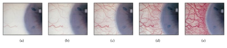
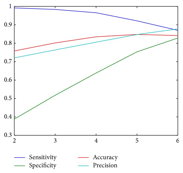
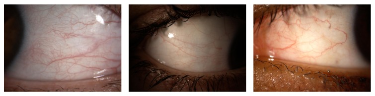
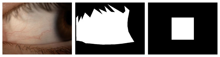
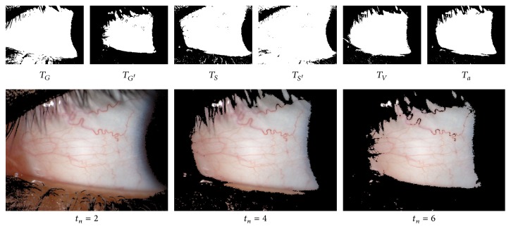
Conjunctival hyperemia or conjunctival redness is a symptom that can be associated with a broad group of ocular diseases. Its levels of severity are represented by standard photographic charts that are visually compared with the patient's eye. This way, the hyperemia diagnosis becomes a nonrepeatable task that depends on the experience of the grader. To solve this problem, we have proposed a computer-aided methodology that comprises three main stages: the segmentation of the conjunctiva, the extraction of features in this region based on colour and the presence of blood vessels, and, finally, the transformation of these features into grading scale values by means of regression techniques. However, the conjunctival segmentation can be slightly inaccurate mainly due to illumination issues. In this work, we analyse the relevance of different features with respect to their location within the conjunctiva in order to delimit a reliable region of interest for the grading. The results show that the automatic procedure behaves like an expert using only a limited region of interest within the conjunctiva.

摘要

结膜充血或结膜发红是一种可与多种眼部疾病相关的症状。其严重程度通过标准摄影图表来表示,这些图表通过视觉与患者的眼睛进行比较。这样,充血诊断就成为一项依赖于分级者经验的不可重复的任务。为了解决这个问题,我们提出了一种计算机辅助方法,该方法包括三个主要阶段:结膜分割、基于颜色和血管存在情况在该区域提取特征,以及最后通过回归技术将这些特征转换为分级量表值。然而,结膜分割可能会稍有不准确,主要是由于光照问题。在这项工作中,我们分析了不同特征相对于它们在结膜内位置的相关性,以便为分级划定一个可靠的感兴趣区域。结果表明,自动程序仅使用结膜内有限的感兴趣区域就能像专家一样发挥作用。

https://cdn.ncbi.nlm.nih.gov/pmc/blobs/ee45/5206783/be2d65b163dd/CMMM2016-3695014.006.jpg
https://cdn.ncbi.nlm.nih.gov/pmc/blobs/ee45/5206783/c24d7c293cc7/CMMM2016-3695014.001.jpg
https://cdn.ncbi.nlm.nih.gov/pmc/blobs/ee45/5206783/48b8be436723/CMMM2016-3695014.002.jpg
https://cdn.ncbi.nlm.nih.gov/pmc/blobs/ee45/5206783/d6283c621053/CMMM2016-3695014.003.jpg
https://cdn.ncbi.nlm.nih.gov/pmc/blobs/ee45/5206783/9633ab1921ba/CMMM2016-3695014.004.jpg
https://cdn.ncbi.nlm.nih.gov/pmc/blobs/ee45/5206783/b478d453606b/CMMM2016-3695014.005.jpg
https://cdn.ncbi.nlm.nih.gov/pmc/blobs/ee45/5206783/be2d65b163dd/CMMM2016-3695014.006.jpg

相似文献

1
Defining the Optimal Region of Interest for Hyperemia Grading in the Bulbar Conjunctiva.确定球结膜充血分级的最佳感兴趣区域
Comput Math Methods Med. 2016;2016:3695014. doi: 10.1155/2016/3695014. Epub 2016 Dec 19.
2
On the development of conjunctival hyperemia computer-assisted diagnosis tools: Influence of feature selection and class imbalance in automatic gradings.关于结膜充血计算机辅助诊断工具的开发:特征选择和类别不平衡对自动分级的影响。
Artif Intell Med. 2016 Jul;71:30-42. doi: 10.1016/j.artmed.2016.06.004. Epub 2016 Jun 30.
3
Hyperemia Analysis Software for Assessment of Conjunctival Hyperemia Severity.充血分析软件用于评估结膜充血严重程度。
Curr Eye Res. 2019 Apr;44(4):376-380. doi: 10.1080/02713683.2018.1554153. Epub 2018 Dec 12.
4
How red is a white eye? Clinical grading of normal conjunctival hyperaemia.白眼有多红?正常结膜充血的临床分级。
Eye (Lond). 2007 May;21(5):633-8. doi: 10.1038/sj.eye.6702295. Epub 2006 Mar 3.
5
Automated hyperemia analysis software: reliability and reproducibility in healthy subjects.自动充血分析软件:健康受试者中的可靠性和可重复性。
Jpn J Ophthalmol. 2012 Jan;56(1):1-7. doi: 10.1007/s10384-011-0107-2. Epub 2011 Dec 1.
6
Development and Evaluation of Semiautomated Quantification of Lissamine Green Staining of the Bulbar Conjunctiva From Digital Images.基于数字图像的球结膜丽丝胺绿染色半自动定量分析方法的开发与评估
JAMA Ophthalmol. 2017 Oct 1;135(10):1078-1085. doi: 10.1001/jamaophthalmol.2017.3346.
7
Sensitivity and reliability of objective image analysis compared to subjective grading of bulbar hyperaemia.与球结膜充血主观分级相比,客观图像分析的敏感性和可靠性。
Br J Ophthalmol. 2007 Nov;91(11):1464-6. doi: 10.1136/bjo.2006.112680. Epub 2007 May 2.
8
Development of automated conjunctival hyperemia analysis software.自动化结膜充血分析软件的开发。
Cornea. 2013 Nov;32 Suppl 1:S52-9. doi: 10.1097/ICO.0b013e3182a18e44.
9
Assessing ocular bulbar redness: a comparison of methods.评估眼球结膜充血:方法比较
Ophthalmic Physiol Opt. 2016 Mar;36(2):132-9. doi: 10.1111/opo.12245. Epub 2015 Nov 17.
10
Temperature of the hyperemic bulbar conjunctiva.充血性球结膜温度。
Curr Eye Res. 1988 Jun;7(6):615-8. doi: 10.3109/02713688809031818.

引用本文的文献

1
Advancing Diagnostics with Semi-Automatic Tear Meniscus Central Area Measurement for Aqueous Deficient Dry Eye Discrimination.通过半自动泪液半月板中央区域测量推进诊断以鉴别水样液缺乏性干眼
Medicina (Kaunas). 2025 Jul 22;61(8):1322. doi: 10.3390/medicina61081322.
2
Toward automated assessment of conjunctival hyperemia: A semisupervised artificial intelligence approach.迈向结膜充血的自动化评估:一种半监督人工智能方法。
Ann N Y Acad Sci. 2025 Sep;1551(1):201-209. doi: 10.1111/nyas.70009. Epub 2025 Aug 6.
3
Conjunctival Bulbar Redness Extraction Pipeline for High-Resolution Ocular Surface Photography.

本文引用的文献

1
Polyphasic analysis of an Azoarcus-Leptothrix-dominated bacterial biofilm developed on stainless steel surface in a gasoline-contaminated hypoxic groundwater.在受汽油污染的缺氧地下水中,对在不锈钢表面形成的以固氮弧菌-纤发菌为主的细菌生物膜进行多相分析。
Environ Sci Pollut Res Int. 2016 May;23(9):9019-35. doi: 10.1007/s11356-016-6128-0. Epub 2016 Jan 29.
2
Analysis of magnetosome chains in magnetotactic bacteria by magnetic measurements and automated image analysis of electron micrographs.通过磁测量和电子显微镜照片的自动图像分析对趋磁细菌中的磁小体链进行分析。
Appl Environ Microbiol. 2013 Dec;79(24):7755-62. doi: 10.1128/AEM.02143-13. Epub 2013 Oct 4.
3
用于高分辨率眼表摄影的结膜球结膜充血提取流程
Transl Vis Sci Technol. 2025 Jan 2;14(1):6. doi: 10.1167/tvst.14.1.6.
4
Meibomian gland secretion quality association with ocular parameters in university students during COVID- 19 restrictions.新冠疫情期间大学生的睑板腺分泌质量与眼部参数的关系。
Int Ophthalmol. 2023 Jul;43(7):2349-2362. doi: 10.1007/s10792-023-02632-2. Epub 2023 Jan 24.
5
Ocular redness - I: Etiology, pathogenesis, and assessment of conjunctival hyperemia.眼部发红 - I:结膜充血的病因、发病机制及评估
Ocul Surf. 2021 Jul;21:134-144. doi: 10.1016/j.jtos.2021.05.003. Epub 2021 May 16.
6
Application of artificial intelligence in anterior segment ophthalmic diseases: diversity and standardization.人工智能在眼前段眼科疾病中的应用:多样性与标准化
Ann Transl Med. 2020 Jun;8(11):714. doi: 10.21037/atm-20-976.
7
Development and Evaluation of Semiautomated Quantification of Lissamine Green Staining of the Bulbar Conjunctiva From Digital Images.基于数字图像的球结膜丽丝胺绿染色半自动定量分析方法的开发与评估
JAMA Ophthalmol. 2017 Oct 1;135(10):1078-1085. doi: 10.1001/jamaophthalmol.2017.3346.
Biosurfactant production by a soil pseudomonas strain growing on polycyclic aromatic hydrocarbons.
土壤假单胞菌菌株在多环芳烃上的生物表面活性剂生产。
Appl Environ Microbiol. 1996 Jun;62(6):1908-12. doi: 10.1128/aem.62.6.1908-1912.1996.
4
Anaerobic degradation of benzene, toluene, ethylbenzene, and xylene compounds by Dechloromonas strain RCB.脱氯单胞菌属菌株RCB对苯、甲苯、乙苯和二甲苯化合物的厌氧降解作用
Appl Environ Microbiol. 2005 Dec;71(12):8649-55. doi: 10.1128/AEM.71.12.8649-8655.2005.
5
Magnetosome formation in prokaryotes.原核生物中磁小体的形成。
Nat Rev Microbiol. 2004 Mar;2(3):217-30. doi: 10.1038/nrmicro842.
6
A review and update of the microbiology of enhanced biological phosphorus removal in wastewater treatment plants.污水处理厂强化生物除磷微生物学的综述与更新
Antonie Van Leeuwenhoek. 2002 Aug;81(1-4):681-91. doi: 10.1023/a:1020538429009.
7
Microbial aspects of anaerobic BTEX degradation.厌氧BTEX降解的微生物学方面
Biomed Environ Sci. 2002 Jun;15(2):130-44.