Nissim Nicole R, O'Shea Andrew M, Bryant Vaughn, Porges Eric C, Cohen Ronald, Woods Adam J
Center for Cognitive Aging and Memory, McKnight Brain Institute, Department of Clinical and Health Psychology, University of FloridaGainesville, FL, USA; Department of Neuroscience, University of FloridaGainesville, FL, USA.
Center for Cognitive Aging and Memory, McKnight Brain Institute, Department of Clinical and Health Psychology, University of Florida Gainesville, FL, USA.
Front Aging Neurosci. 2017 Jan 4;8:328. doi: 10.3389/fnagi.2016.00328. eCollection 2016.
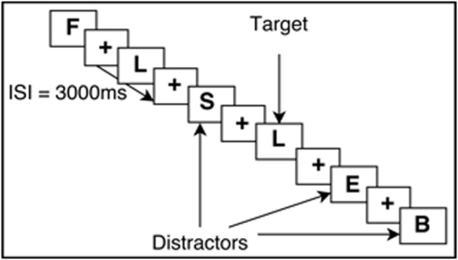
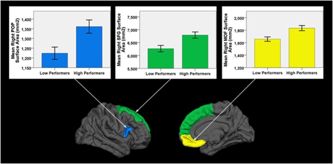
Working memory is an executive memory process that allows transitional information to be held and manipulated temporarily in memory stores before being forgotten or encoded into long-term memory. Working memory is necessary for everyday decision-making and problem solving, making it a fundamental process in the daily lives of older adults. Working memory relies heavily on frontal lobe structures and is known to decline with age. The current study aimed to determine the neural correlates of decreased working memory performance in the frontal lobes by comparing cortical thickness and cortical surface area from two demographically matched groups of healthy older adults, free from cognitive impairment, with high versus low N-Back working memory performance ( = 56; average age = 70.29 ± 10.64). High-resolution structural T1-weighted images (1 mm isotropic voxels) were obtained on a 3T Philips MRI scanner. When compared to high performers, low performers exhibited significantly decreased cortical surface area in three frontal lobe regions lateralized to the right hemisphere: medial orbital frontal gyrus, inferior frontal gyrus, and superior frontal gyrus (FDR < 0.05). There were no significant differences in cortical thickness between groups, a proxy for neurodegenerative tissue loss. Our results suggest that decreases in cortical surface area (a proxy for brain structural integrity) in right frontal regions may underlie age-related decline of working memory function.
工作记忆是一种执行性记忆过程,它允许过渡性信息在被遗忘或编码到长期记忆之前,在记忆存储中被暂时保存和处理。工作记忆对于日常决策和问题解决至关重要,使其成为老年人日常生活中的一个基本过程。工作记忆严重依赖额叶结构,并且已知会随着年龄增长而下降。当前的研究旨在通过比较两组在人口统计学上匹配的、无认知障碍的健康老年人的皮质厚度和皮质表面积,来确定额叶中工作记忆表现下降的神经关联,这两组老年人的N-回溯工作记忆表现分别为高和低(n = 56;平均年龄 = 70.29 ± 10.64)。在一台3T飞利浦MRI扫描仪上获取了高分辨率结构T1加权图像(1毫米各向同性体素)。与高表现者相比,低表现者在右侧半球的三个额叶区域表现出明显减小的皮质表面积:眶额内侧回、额下回和额上回(FDR < 0.05)。两组之间的皮质厚度没有显著差异,皮质厚度可作为神经退行性组织损失的一个指标。我们的结果表明,右侧额叶区域皮质表面积的减小(可作为脑结构完整性的一个指标)可能是与年龄相关的工作记忆功能衰退的基础。