Gesnik Marc, Blaize Kevin, Deffieux Thomas, Gennisson Jean-Luc, Sahel José-Alain, Fink Mathias, Picaud Serge, Tanter Mickaël
Institut Langevin, ESPCI Paris, PSL Research University, CNRS UMR 7587, INSERM U979, 75012 Paris, France.
Institut de la Vision, Sorbonne Universités UPMC, University of Paris 06, INSERM UMR_S 968, CNRS UMR 7210, 75012 Paris, France.
Neuroimage. 2017 Apr 1;149:267-274. doi: 10.1016/j.neuroimage.2017.01.071. Epub 2017 Feb 3.
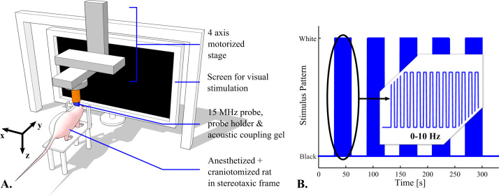
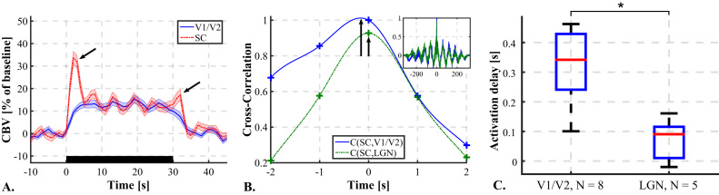
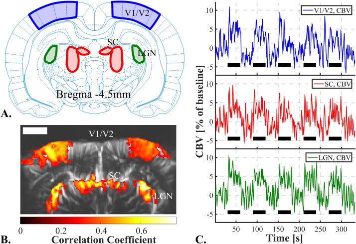
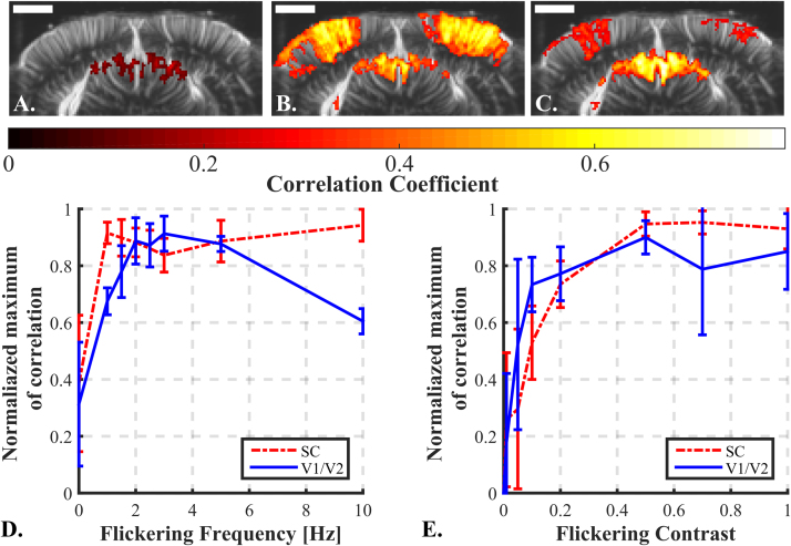
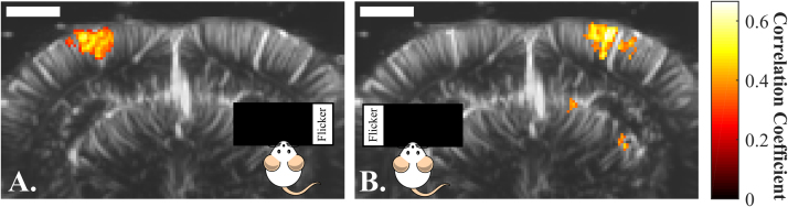
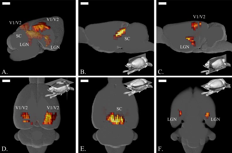
3D functional imaging of the whole brain activity during visual task is a challenging task in rodents due to the complex tri-dimensional shape of involved brain regions and the fine spatial and temporal resolutions required to reveal the visual tract. By coupling functional ultrasound (fUS) imaging with a translational motorized stage and an episodic visual stimulation device, we managed to accurately map and to recover the activity of the visual cortices, the Superior Colliculus (SC) and the Lateral Geniculate Nuclei (LGN) in 3D. Cerebral Blood Volume (CBV) responses during visual stimuli were found to be highly correlated with the visual stimulus time profile in visual cortices (r=0.6), SC (r=0.7) and LGN (r=0.7). These responses were found dependent on flickering frequency and contrast, and optimal stimulus parameters for largest CBV increases were obtained. In particular, increasing the flickering frequency higher than 7Hz revealed a decrease of visual cortices response while the SC response was preserved. Finally, cross-correlation between CBV signals exhibited significant delays (d=0.35s +/-0.1s) between blood volume response in SC and visual cortices in response to our visual stimulus. These results emphasize the interest of fUS imaging as a whole brain neuroimaging modality for brain vision studies in rodent models.
在啮齿动物中,由于所涉及脑区复杂的三维形状以及揭示视觉通路所需的精细空间和时间分辨率,对视觉任务期间全脑活动进行三维功能成像颇具挑战性。通过将功能超声(fUS)成像与平移电动平台和间歇性视觉刺激装置相结合,我们成功地在三维空间中精确绘制并恢复了视觉皮层、上丘(SC)和外侧膝状体(LGN)的活动。研究发现,视觉刺激期间的脑血容量(CBV)反应与视觉皮层(r = 0.6)、SC(r = 0.7)和LGN(r = 0.7)中的视觉刺激时间曲线高度相关。这些反应取决于闪烁频率和对比度,并获得了使CBV最大增加的最佳刺激参数。特别是,将闪烁频率提高到7Hz以上会导致视觉皮层反应降低,而上丘反应则得以保留。最后,CBV信号之间的互相关显示,在我们的视觉刺激下,SC中的血容量反应与视觉皮层之间存在显著延迟(d = 0.35秒±0.1秒)。这些结果强调了fUS成像作为一种全脑神经成像方式在啮齿动物模型脑视觉研究中的价值。