Keuken M C, Bazin P-L, Backhouse K, Beekhuizen S, Himmer L, Kandola A, Lafeber J J, Prochazkova L, Trutti A, Schäfer A, Turner R, Forstmann B U
Integrative Model-based Cognitive Neuroscience Research Unit, University of Amsterdam, Amsterdam, The Netherlands.
Netherlands Institute for Neuroscience, an Institute of the Royal Netherlands Academy of Arts and Sciences, Amsterdam, The Netherlands.
Brain Struct Funct. 2017 Aug;222(6):2487-2505. doi: 10.1007/s00429-016-1352-4. Epub 2017 Feb 6.
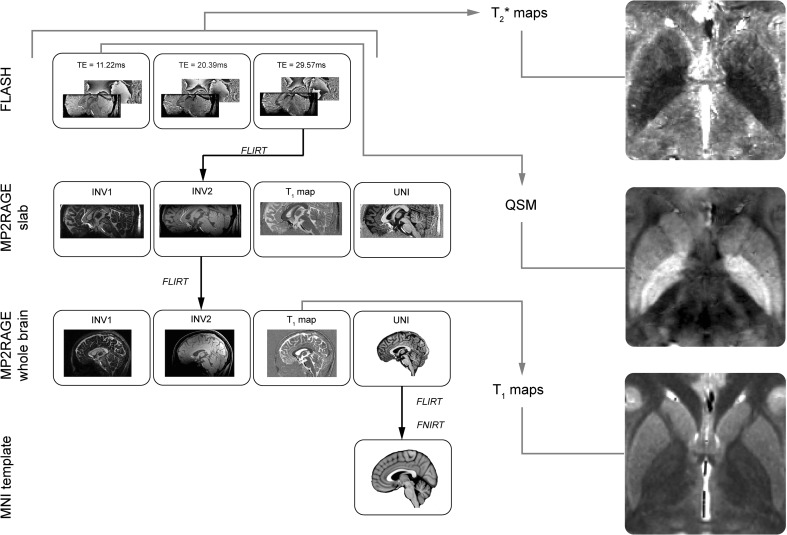
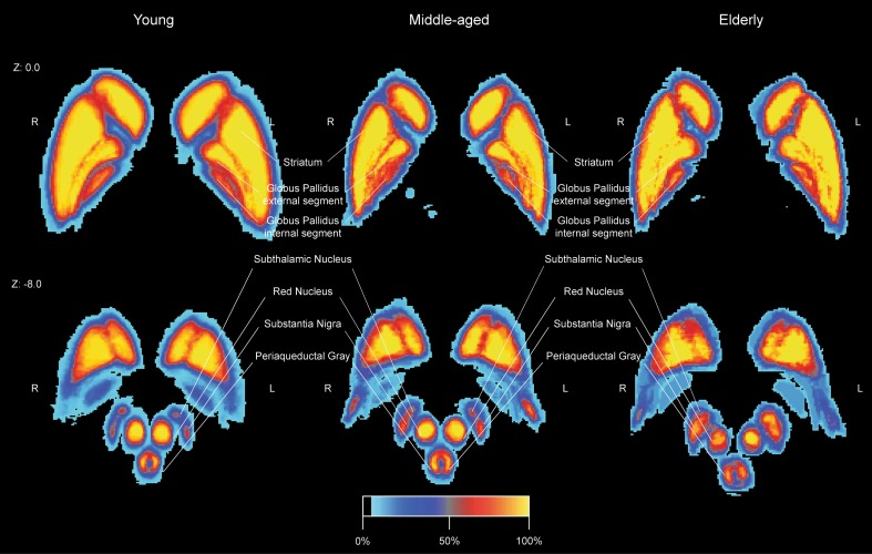
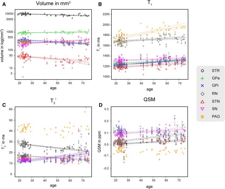
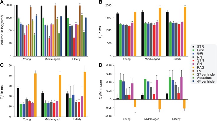
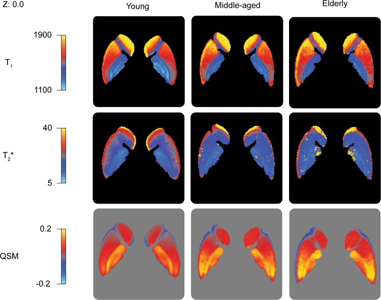
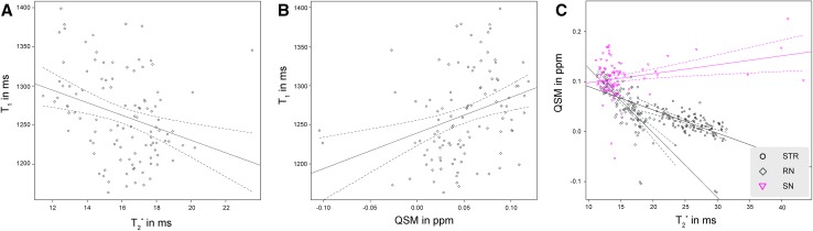
The aging brain undergoes several anatomical changes that can be measured with Magnetic Resonance Imaging (MRI). Early studies using lower field strengths have assessed changes in tissue properties mainly qualitatively, using [Formula: see text]- or [Formula: see text]- weighted images to provide image contrast. With the development of higher field strengths (7 T and above) and more advanced MRI contrasts, quantitative measures can be acquired even of small subcortical structures. This study investigates volumetric, spatial, and quantitative MRI parameter changes associated with healthy aging in a range of subcortical nuclei, including the basal ganglia, red nucleus, and the periaqueductal grey. The results show that aging has a heterogenous effects across regions. Across the subcortical areas an increase of [Formula: see text] values is observed, most likely indicating a loss of myelin. Only for a number of areas, a decrease of [Formula: see text] and increase of QSM is found, indicating an increase of iron. Aging also results in a location shift for a number of structures indicating the need for visualization of the anatomy of individual brains.
衰老的大脑会经历一些可通过磁共振成像(MRI)测量的解剖学变化。早期使用较低场强的研究主要通过定性评估组织特性,使用T1加权或T2加权图像来提供图像对比度。随着更高场强(7T及以上)和更先进的MRI对比度的发展,即使是小的皮质下结构也可以获得定量测量结果。本研究调查了一系列皮质下核团(包括基底神经节、红核和导水管周围灰质)中与健康衰老相关的体积、空间和定量MRI参数变化。结果表明,衰老对不同区域有不同的影响。在整个皮质下区域观察到T2*值增加,最有可能表明髓鞘丢失。仅在一些区域发现T2值降低和QSM增加,表明铁含量增加。衰老还导致一些结构的位置发生偏移,这表明需要对个体大脑的解剖结构进行可视化。