Kashyap Sriranga, Ivanov Dimo, Havlicek Martin, Huber Laurentius, Poser Benedikt A, Uludağ Kâmil
Department of Cognitive Neuroscience, Faculty of Psychology and Neuroscience, Maastricht University, Maastricht, The Netherlands.
Maastricht Brain Imaging Centre (M-BIC), Maastricht University, Maastricht, The Netherlands.
PLoS One. 2021 Apr 26;16(4):e0250504. doi: 10.1371/journal.pone.0250504. eCollection 2021.
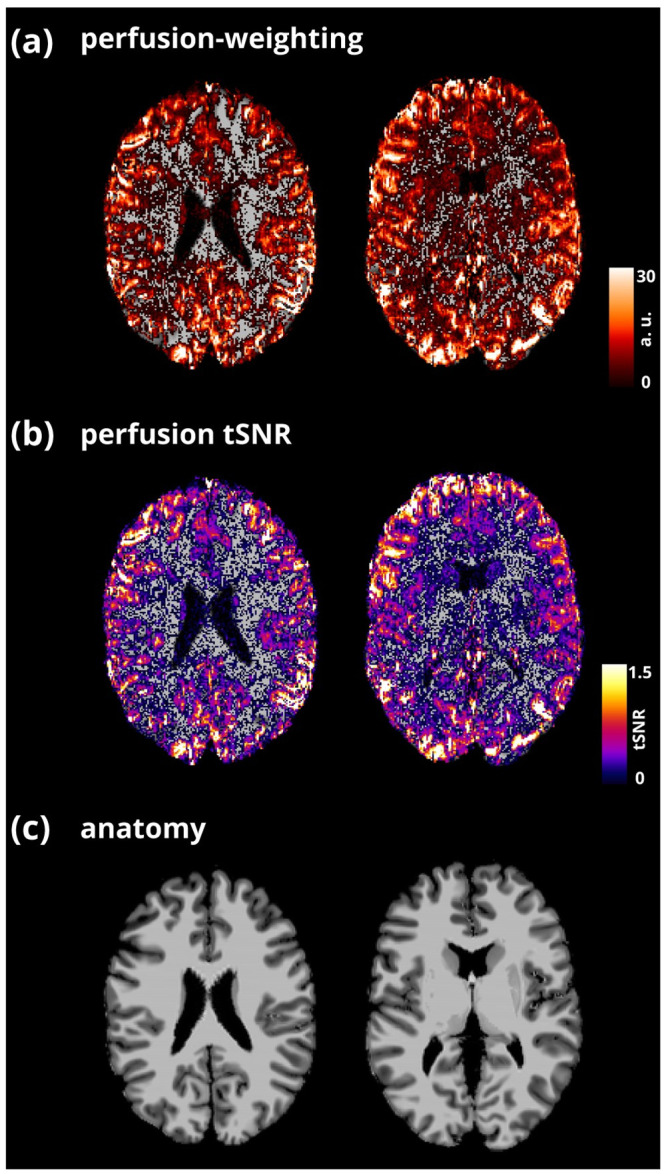
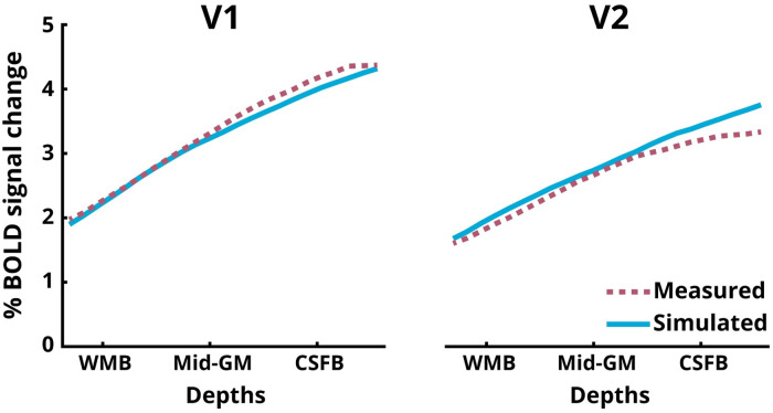
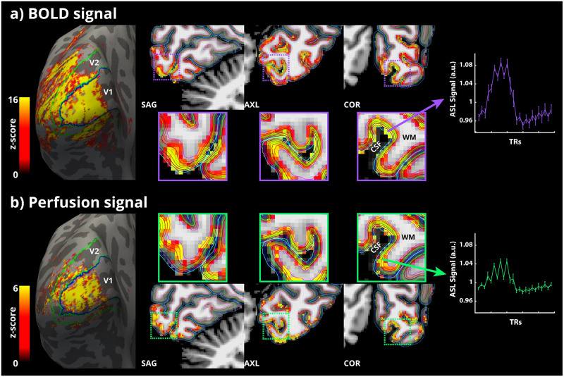
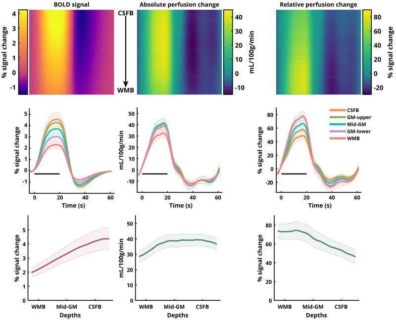
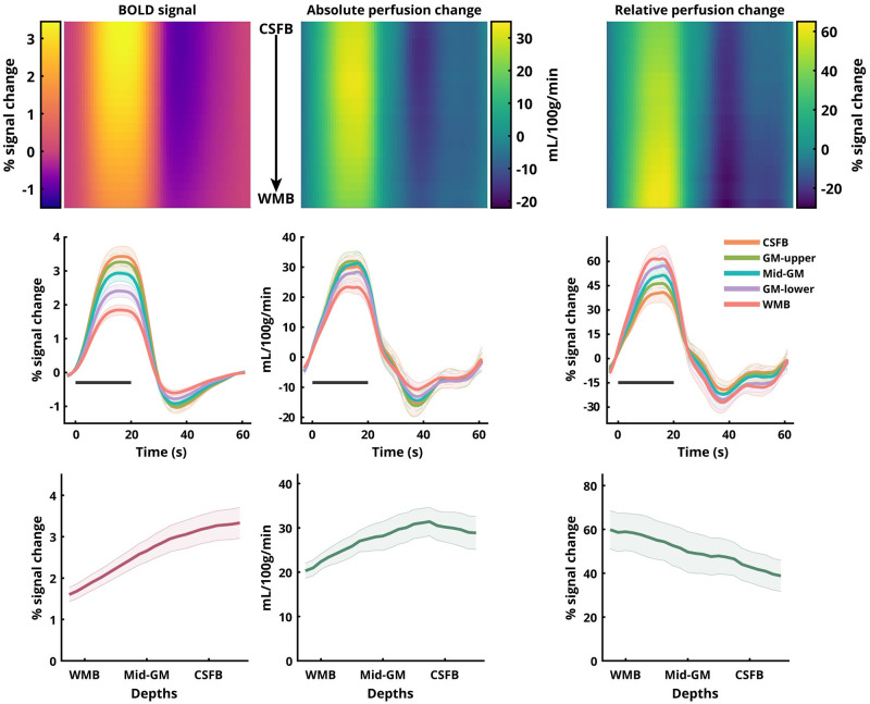
Laminar fMRI at ultra-high magnetic field strength is typically carried out using the Blood Oxygenation Level-Dependent (BOLD) contrast. Despite its unrivalled sensitivity to detecting activation, the BOLD contrast is limited in its spatial specificity due to signals stemming from intra-cortical ascending and pial veins. Alternatively, regional changes in perfusion (i.e., cerebral blood flow through tissue) are colocalised to neuronal activation, which can be non-invasively measured using Arterial Spin Labelling (ASL) MRI. In addition, ASL provides a quantitative marker of neuronal activation in terms of perfusion signal, which is simultaneously acquired along with the BOLD signal. However, ASL for laminar imaging is challenging due to the lower SNR of the perfusion signal and higher RF power deposition i.e., specific absorption rate (SAR) of ASL sequences. In the present study, we present for the first time in humans, isotropic sub-millimetre spatial resolution functional perfusion images using Flow-sensitive Alternating Inversion Recovery (FAIR) ASL with a 3D-EPI readout at 7 T. We show that robust statistical activation maps can be obtained with perfusion-weighting in a single session. We observed the characteristic BOLD amplitude increase towards the superficial laminae, and, in apparent discrepancy, the relative perfusion profile shows a decrease of the amplitude and the absolute perfusion profile a much smaller increase towards the cortical surface. Considering the draining vein effect on the BOLD signal using model-based spatial "convolution", we show that the empirically measured perfusion and BOLD profiles are, in fact, consistent with each other. This study demonstrates that laminar perfusion fMRI in humans is feasible at 7 T and that caution must be exercised when interpreting BOLD signal laminar profiles as direct representation of the cortical distribution of neuronal activity.
超高磁场强度下的层流功能磁共振成像通常使用血氧水平依赖(BOLD)对比进行。尽管其在检测激活方面具有无与伦比的灵敏度,但由于来自皮质内升支静脉和软脑膜静脉的信号,BOLD对比在空间特异性方面存在局限性。或者,灌注的区域变化(即通过组织的脑血流量)与神经元激活共定位,这可以使用动脉自旋标记(ASL)磁共振成像进行无创测量。此外,ASL根据灌注信号提供神经元激活的定量标记,该信号与BOLD信号同时采集。然而,由于灌注信号的信噪比低以及ASL序列的射频功率沉积较高,即比吸收率(SAR),用于层流成像的ASL具有挑战性。在本研究中,我们首次在人体中展示了使用具有3D-EPI读出的血流敏感交替反转恢复(FAIR)ASL在7T时获得的各向同性亚毫米空间分辨率功能灌注图像。我们表明,在单次采集中通过灌注加权可以获得强大的统计激活图。我们观察到向浅表层的特征性BOLD幅度增加,并且明显矛盾的是,相对灌注曲线显示幅度降低,而绝对灌注曲线显示向皮质表面的增加要小得多。使用基于模型的空间“卷积”考虑引流静脉对BOLD信号的影响,我们表明经验测量的灌注和BOLD曲线实际上彼此一致。这项研究表明,人体层流灌注功能磁共振成像在7T时是可行的,并且在将BOLD信号层流曲线解释为神经元活动皮质分布的直接表示时必须谨慎。