Atta-Fosu Thomas, Guo Weihong, Jeter Dana, Mizutani Claudia M, Stopczynski Nathan, Sousa-Neves Rui
Department of Mathematics, Applied Mathematics and Statistics, Case Western Reserve University, 10900 Euclid Avenue, Cleveland, OH 44106, USA.
Department of Biology, Case Western Reserve University, 10900 Euclid Avenue, Cleveland, OH 44106, USA.
J Imaging. 2016 Dec;2(4). doi: 10.3390/jimaging2040031. Epub 2016 Nov 5.
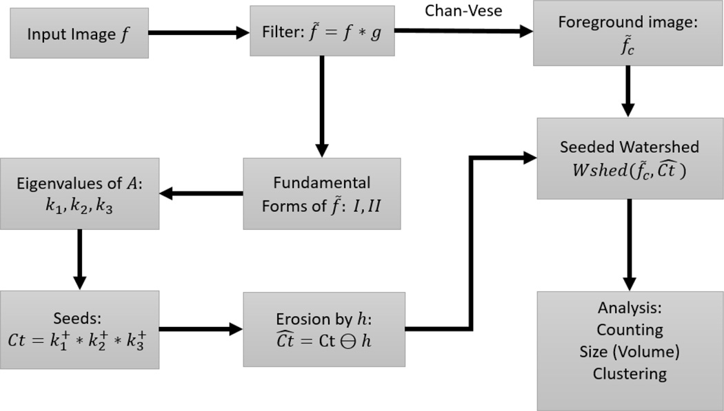
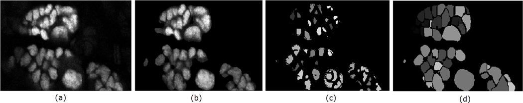
Image segmentation is an important process that separates objects from the background and also from each other. Applied to cells, the results can be used for cell counting which is very important in medical diagnosis and treatment, and biological research that is often used by scientists and medical practitioners. Segmenting 3D confocal microscopy images containing cells of different shapes and sizes is still challenging as the nuclei are closely packed. The watershed transform provides an efficient tool in segmenting such nuclei provided a reasonable set of markers can be found in the image. In the presence of low-contrast variation or excessive noise in the given image, the watershed transform leads to over-segmentation (a single object is overly split into multiple objects). The traditional watershed uses the local minima of the input image and will characteristically find multiple minima in one object unless they are specified (marker-controlled watershed). An alternative to using the local minima is by a supervised technique called seeded watershed, which supplies single seeds to replace the minima for the objects. Consequently, the accuracy of a seeded watershed algorithm relies on the accuracy of the predefined seeds. In this paper, we present a segmentation approach based on the geometric morphological properties of the 'landscape' using curvatures. The curvatures are computed as the eigenvalues of the Shape matrix, producing accurate seeds that also inherit the original shape of their respective cells. We compare with some popular approaches and show the advantage of the proposed method.
图像分割是一个重要的过程,它将物体与背景以及物体之间相互分离。应用于细胞时,其结果可用于细胞计数,这在医学诊断和治疗以及生物研究中非常重要,而生物研究常被科学家和医学从业者所采用。对包含不同形状和大小细胞的三维共聚焦显微镜图像进行分割仍然具有挑战性,因为细胞核紧密排列。分水岭变换提供了一种有效的工具来分割此类细胞核,前提是能在图像中找到一组合理的标记。在给定图像存在低对比度变化或过多噪声的情况下,分水岭变换会导致过分割(单个物体被过度分割成多个物体)。传统的分水岭变换使用输入图像的局部最小值,并且通常会在一个物体中找到多个最小值,除非这些最小值被指定(标记控制的分水岭变换)。使用局部最小值的一种替代方法是一种称为种子分水岭的监督技术,它提供单个种子来替代物体的最小值。因此,种子分水岭算法的准确性依赖于预定义种子的准确性。在本文中,我们提出了一种基于使用曲率的“地形”几何形态属性的分割方法。曲率被计算为形状矩阵的特征值,从而产生准确的种子,这些种子还继承了各自细胞的原始形状。我们与一些流行的方法进行比较,并展示了所提出方法的优势。