• 文献检索
  • 文档翻译
  • 深度研究
  • 学术资讯
  • Suppr Zotero 插件Zotero 插件
  • 邀请有礼
  • 套餐&价格
  • 历史记录
应用&插件
Suppr Zotero 插件Zotero 插件浏览器插件Mac 客户端Windows 客户端微信小程序
定价
高级版会员购买积分包购买API积分包
服务
文献检索文档翻译深度研究API 文档MCP 服务
关于我们
关于 Suppr公司介绍联系我们用户协议隐私条款
关注我们

Suppr 超能文献

核心技术专利:CN118964589B侵权必究
粤ICP备2023148730 号-1Suppr @ 2026

文献检索

告别复杂PubMed语法,用中文像聊天一样搜索,搜遍4000万医学文献。AI智能推荐,让科研检索更轻松。

立即免费搜索

文件翻译

保留排版,准确专业,支持PDF/Word/PPT等文件格式,支持 12+语言互译。

免费翻译文档

深度研究

AI帮你快速写综述,25分钟生成高质量综述,智能提取关键信息,辅助科研写作。

立即免费体验

基于K均值聚类和分水岭算法的图形图像区域提取

Graphical Image Region Extraction with K-Means Clustering and Watershed.

作者信息

Jardim Sandra, António João, Mora Carlos

机构信息

Smart Cities Research Center, Polytechnic Institute of Tomar, 2300-313 Tomar, Portugal.

Techframe-Information Systems, SA, 2785-338 São Domingos de Rana, Portugal.

出版信息

J Imaging. 2022 Jun 8;8(6):163. doi: 10.3390/jimaging8060163.

DOI:10.3390/jimaging8060163
PMID:35735962
原文链接:https://pmc.ncbi.nlm.nih.gov/articles/PMC9224791/
Abstract

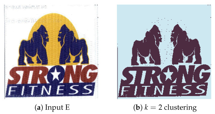
With a wide range of applications, image segmentation is a complex and difficult preprocessing step that plays an important role in automatic visual systems, which accuracy impacts, not only on segmentation results, but directly affects the effectiveness of the follow-up tasks. Despite the many advances achieved in the last decades, image segmentation remains a challenging problem, particularly, the segmenting of color images due to the diverse inhomogeneities of color, textures and shapes present in the descriptive features of the images. In trademark graphic images segmentation, beyond these difficulties, we must also take into account the high noise and low resolution, which are often present. Trademark graphic images can also be very heterogeneous with regard to the elements that make them up, which can be overlapping and with varying lighting conditions. Due to the immense variation encountered in corporate logos and trademark graphic images, it is often difficult to select a single method for extracting relevant image regions in a way that produces satisfactory results. Many of the hybrid approaches that integrate the Watershed and K-Means algorithms involve processing very high quality and visually similar images, such as medical images, meaning that either approach can be tweaked to work on images that follow a certain pattern. Trademark images are totally different from each other and are usually fully colored. Our system solves this difficulty given it is a generalized implementation designed to work in most scenarios, through the use of customizable parameters and completely unbiased for an image type. In this paper, we propose a hybrid approach to Image Region Extraction that focuses on automated region proposal and segmentation techniques. In particular, we analyze popular techniques such as K-Means Clustering and Watershedding and their effectiveness when deployed in a hybrid environment to be applied to a highly variable dataset. The proposed system consists of a multi-stage algorithm that takes as input an RGB image and produces multiple outputs, corresponding to the extracted regions. After preprocessing steps, a K-Means function with random initial centroids and a user-defined value for is executed over the RGB image, generating a gray-scale segmented image, to which a threshold method is applied to generate a binary mask, containing the necessary information to generate a distance map. Then, the Watershed function is performed over the distance map, using the markers defined by the Connected Component Analysis function that labels regions on 8-way pixel connectivity, ensuring that all regions are correctly found. Finally, individual objects are labelled for extraction through a contour method, based on border following. The achieved results show adequate region extraction capabilities when processing graphical images from different datasets, where the system correctly distinguishes the most relevant visual elements of images with minimal tweaking.

摘要

图像分割具有广泛的应用,是一个复杂且困难的预处理步骤,在自动视觉系统中起着重要作用,其准确性不仅影响分割结果,还直接影响后续任务的有效性。尽管在过去几十年中取得了许多进展,但图像分割仍然是一个具有挑战性的问题,特别是彩色图像的分割,因为图像的描述特征中存在颜色、纹理和形状的各种不均匀性。在商标图形图像分割中,除了这些困难之外,我们还必须考虑经常出现的高噪声和低分辨率。商标图形图像在构成它们的元素方面也可能非常不同,这些元素可能相互重叠且光照条件各异。由于企业标志和商标图形图像中存在巨大差异,通常很难选择一种单一方法来以产生满意结果的方式提取相关图像区域。许多集成了分水岭算法和K均值算法的混合方法涉及处理非常高质量且视觉上相似的图像,例如医学图像,这意味着可以调整任何一种方法以处理遵循特定模式的图像。商标图像彼此完全不同,通常是全彩色的。我们的系统解决了这个难题,因为它是一个通用实现,旨在通过使用可定制参数并对图像类型完全无偏地在大多数场景中工作。在本文中,我们提出了一种用于图像区域提取的混合方法,该方法侧重于自动区域提议和分割技术。特别是,我们分析了诸如K均值聚类和分水岭算法等流行技术,以及它们在混合环境中部署以应用于高度可变数据集时的有效性。所提出的系统由一个多阶段算法组成,该算法将RGB图像作为输入并产生多个输出,对应于提取的区域。在预处理步骤之后,对RGB图像执行具有随机初始质心和用户定义的 值的K均值函数,生成一个灰度分割图像,对其应用阈值方法以生成一个二进制掩码,该掩码包含生成距离图所需的信息。然后,使用由连通分量分析函数定义的标记在距离图上执行分水岭函数,该函数基于8向像素连通性标记区域,确保正确找到所有区域。最后,通过基于边界跟踪的轮廓方法对单个对象进行标记以进行提取。在处理来自不同数据集的图形图像时,所取得的结果显示出足够的区域提取能力,该系统在进行最少调整的情况下能够正确区分图像中最相关的视觉元素。

https://cdn.ncbi.nlm.nih.gov/pmc/blobs/9448/9224791/7cfc36655da8/jimaging-08-00163-g025.jpg
https://cdn.ncbi.nlm.nih.gov/pmc/blobs/9448/9224791/c231763f3508/jimaging-08-00163-g001.jpg
https://cdn.ncbi.nlm.nih.gov/pmc/blobs/9448/9224791/11cb88c8dc13/jimaging-08-00163-g002.jpg
https://cdn.ncbi.nlm.nih.gov/pmc/blobs/9448/9224791/7a30ddaaea38/jimaging-08-00163-g003.jpg
https://cdn.ncbi.nlm.nih.gov/pmc/blobs/9448/9224791/6eed3d4fdfe6/jimaging-08-00163-g004.jpg
https://cdn.ncbi.nlm.nih.gov/pmc/blobs/9448/9224791/567854e83dc5/jimaging-08-00163-g005.jpg
https://cdn.ncbi.nlm.nih.gov/pmc/blobs/9448/9224791/0e788511c9b2/jimaging-08-00163-g006.jpg
https://cdn.ncbi.nlm.nih.gov/pmc/blobs/9448/9224791/b8f51b9b8fe8/jimaging-08-00163-g007.jpg
https://cdn.ncbi.nlm.nih.gov/pmc/blobs/9448/9224791/2b97c8eefcc0/jimaging-08-00163-g008.jpg
https://cdn.ncbi.nlm.nih.gov/pmc/blobs/9448/9224791/cf32203fd103/jimaging-08-00163-g009.jpg
https://cdn.ncbi.nlm.nih.gov/pmc/blobs/9448/9224791/02c744f2e83e/jimaging-08-00163-g010.jpg
https://cdn.ncbi.nlm.nih.gov/pmc/blobs/9448/9224791/8b040046d7d9/jimaging-08-00163-g011.jpg
https://cdn.ncbi.nlm.nih.gov/pmc/blobs/9448/9224791/d7bd5a8a0e60/jimaging-08-00163-g012.jpg
https://cdn.ncbi.nlm.nih.gov/pmc/blobs/9448/9224791/7ec49bd7dbc3/jimaging-08-00163-g013.jpg
https://cdn.ncbi.nlm.nih.gov/pmc/blobs/9448/9224791/81f33c8e5a0f/jimaging-08-00163-g014.jpg
https://cdn.ncbi.nlm.nih.gov/pmc/blobs/9448/9224791/c4d92cc77859/jimaging-08-00163-g015.jpg
https://cdn.ncbi.nlm.nih.gov/pmc/blobs/9448/9224791/a5a3a949295f/jimaging-08-00163-g016.jpg
https://cdn.ncbi.nlm.nih.gov/pmc/blobs/9448/9224791/7af02db7c2da/jimaging-08-00163-g017.jpg
https://cdn.ncbi.nlm.nih.gov/pmc/blobs/9448/9224791/16bb4b6f4ae9/jimaging-08-00163-g018.jpg
https://cdn.ncbi.nlm.nih.gov/pmc/blobs/9448/9224791/e9fe6129d318/jimaging-08-00163-g019.jpg
https://cdn.ncbi.nlm.nih.gov/pmc/blobs/9448/9224791/21a641fa5ed3/jimaging-08-00163-g020.jpg
https://cdn.ncbi.nlm.nih.gov/pmc/blobs/9448/9224791/c74a999fb0bc/jimaging-08-00163-g021.jpg
https://cdn.ncbi.nlm.nih.gov/pmc/blobs/9448/9224791/b7bb48b37733/jimaging-08-00163-g022.jpg
https://cdn.ncbi.nlm.nih.gov/pmc/blobs/9448/9224791/4f1bf1ede3f5/jimaging-08-00163-g023.jpg
https://cdn.ncbi.nlm.nih.gov/pmc/blobs/9448/9224791/d44b7f49a370/jimaging-08-00163-g024.jpg
https://cdn.ncbi.nlm.nih.gov/pmc/blobs/9448/9224791/7cfc36655da8/jimaging-08-00163-g025.jpg

相似文献

1
Graphical Image Region Extraction with K-Means Clustering and Watershed.基于K均值聚类和分水岭算法的图形图像区域提取
J Imaging. 2022 Jun 8;8(6):163. doi: 10.3390/jimaging8060163.
2
Bone Region Segmentation in Medical Images Based on Improved Watershed Algorithm.基于改进分水岭算法的医学图像骨骼区域分割。
Comput Intell Neurosci. 2022 Mar 24;2022:3975853. doi: 10.1155/2022/3975853. eCollection 2022.
3
Unsupervised color image segmentation: A case of RGB histogram based K-means clustering initialization.无监督彩色图像分割:基于 RGB 直方图的 K-均值聚类初始化案例。
PLoS One. 2020 Oct 22;15(10):e0240015. doi: 10.1371/journal.pone.0240015. eCollection 2020.
4
Split and Merge Watershed: a two-step method for cell segmentation in fluorescence microscopy images.分裂与合并分水岭算法:一种用于荧光显微镜图像中细胞分割的两步法。
Biomed Signal Process Control. 2019 Aug;53. doi: 10.1016/j.bspc.2019.101575. Epub 2019 Jun 4.
5
FogBank: a single cell segmentation across multiple cell lines and image modalities.FogBank:跨多种细胞系和图像模态的单细胞分割
BMC Bioinformatics. 2014 Dec 30;15(1):431. doi: 10.1186/s12859-014-0431-x.
6
Folic acid supplementation and malaria susceptibility and severity among people taking antifolate antimalarial drugs in endemic areas.在流行地区,服用抗叶酸抗疟药物的人群中,叶酸补充剂与疟疾易感性和严重程度的关系。
Cochrane Database Syst Rev. 2022 Feb 1;2(2022):CD014217. doi: 10.1002/14651858.CD014217.
7
Partitioning histopathological images: an integrated framework for supervised color-texture segmentation and cell splitting.分割组织病理学图像:一种用于监督的颜色-纹理分割和细胞分裂的集成框架。
IEEE Trans Med Imaging. 2011 Sep;30(9):1661-77. doi: 10.1109/TMI.2011.2141674. Epub 2011 Apr 11.
8
Building roof segmentation from aerial images using a lineand region-based watershed segmentation technique.使用基于线条和区域的分水岭分割技术从航空图像中进行建筑物屋顶分割。
Sensors (Basel). 2015 Feb 2;15(2):3172-203. doi: 10.3390/s150203172.
9
An unsupervised automatic segmentation algorithm for breast tissue classification of dedicated breast computed tomography images.一种用于专用乳腺 CT 图像的乳腺组织分类的无监督自动分割算法。
Med Phys. 2018 Jun;45(6):2542-2559. doi: 10.1002/mp.12920. Epub 2018 May 9.
10
A novel method for segmenting brain tumor using modified watershed algorithm in MRI image with FPGA.使用 FPGA 在 MRI 图像中使用改进的分水岭算法对脑肿瘤进行分割的新方法。
Biosystems. 2020 Dec;198:104226. doi: 10.1016/j.biosystems.2020.104226. Epub 2020 Aug 27.

引用本文的文献

1
DarwinGSE: Towards better image retrieval systems for intellectual property datasets.达尔文 GSE:为知识产权数据集建立更好的图像检索系统。
PLoS One. 2024 Jul 1;19(7):e0304915. doi: 10.1371/journal.pone.0304915. eCollection 2024.
2
Developments in Image Processing Using Deep Learning and Reinforcement Learning.利用深度学习和强化学习的图像处理进展
J Imaging. 2023 Sep 30;9(10):207. doi: 10.3390/jimaging9100207.
3
Efficient Approach to Color Image Segmentation Based on Multilevel Thresholding Using EMO Algorithm by Considering Spatial Contextual Information.

本文引用的文献

1
A Review of Watershed Implementations for Segmentation of Volumetric Images.用于体积图像分割的分水岭算法实现综述
J Imaging. 2022 Apr 26;8(5):127. doi: 10.3390/jimaging8050127.
2
Bone Region Segmentation in Medical Images Based on Improved Watershed Algorithm.基于改进分水岭算法的医学图像骨骼区域分割。
Comput Intell Neurosci. 2022 Mar 24;2022:3975853. doi: 10.1155/2022/3975853. eCollection 2022.
3
Boundary-aware context neural network for medical image segmentation.边界感知上下文神经网络在医学图像分割中的应用。
基于多阈值分割并考虑空间上下文信息的高效彩色图像分割方法——使用EMO算法
J Imaging. 2023 Mar 23;9(4):74. doi: 10.3390/jimaging9040074.
4
Multiclass Level-Set Segmentation of Rust and Coating Damages in Images of Metal Structures.金属结构图像中锈蚀与涂层损伤的多类水平集分割
Sensors (Basel). 2022 Oct 7;22(19):7600. doi: 10.3390/s22197600.
5
A Novel Trademark Image Retrieval System Based on Multi-Feature Extraction and Deep Networks.一种基于多特征提取和深度网络的新型商标图像检索系统。
J Imaging. 2022 Sep 2;8(9):238. doi: 10.3390/jimaging8090238.
6
Using DeepLabCut as a Real-Time and Markerless Tool for Cardiac Physiology Assessment in Zebrafish.使用DeepLabCut作为一种用于斑马鱼心脏生理学评估的实时无标记工具。
Biology (Basel). 2022 Aug 21;11(8):1243. doi: 10.3390/biology11081243.
Med Image Anal. 2022 May;78:102395. doi: 10.1016/j.media.2022.102395. Epub 2022 Feb 14.
4
Region-Based CNN for Anomaly Detection in PV Power Plants Using Aerial Imagery.基于区域的卷积神经网络在使用航空图像的光伏电站异常检测中的应用。
Sensors (Basel). 2022 Feb 7;22(3):1244. doi: 10.3390/s22031244.
5
Brain tissue segmentation via non-local fuzzy c-means clustering combined with Markov random field.基于非局部模糊 C 均值聚类与马尔可夫随机场的脑组织分割。
Math Biosci Eng. 2022 Jan;19(2):1891-1908. doi: 10.3934/mbe.2022089. Epub 2021 Dec 20.
6
Intelligent Semantic Segmentation for Self-Driving Vehicles Using Deep Learning.基于深度学习的自动驾驶车辆智能语义分割。
Comput Intell Neurosci. 2022 Jan 17;2022:6390260. doi: 10.1155/2022/6390260. eCollection 2022.
7
Validation of watershed-based segmentation of the cartilage surface from sequential CT arthrography scans.基于分水岭算法的关节造影CT序列扫描软骨表面分割的验证
Quant Imaging Med Surg. 2022 Jan;12(1):1-14. doi: 10.21037/qims-20-1062.
8
Lesion segmentation in breast ultrasound images using the optimized marked watershed method.基于优化标记分水岭算法的乳腺超声图像病灶分割。
Biomed Eng Online. 2021 Jun 7;20(1):57. doi: 10.1186/s12938-021-00891-7.
9
Detection of early decay on citrus using LW-NIR hyperspectral reflectance imaging coupled with two-band ratio and improved watershed segmentation algorithm.利用 LW-NIR 高光谱反射成像技术结合双波段比值和改进的分水岭分割算法检测柑橘早期衰变。
Food Chem. 2021 Oct 30;360:130077. doi: 10.1016/j.foodchem.2021.130077. Epub 2021 May 12.
10
A comprehensive survey of image segmentation: clustering methods, performance parameters, and benchmark datasets.图像分割的全面综述:聚类方法、性能参数和基准数据集。
Multimed Tools Appl. 2022;81(24):35001-35026. doi: 10.1007/s11042-021-10594-9. Epub 2021 Feb 9.