de Barros Garcia Jose Mauricio Botto, Isaac David Leonardo Cruvinel, Avila Marcos
Federal University of Goias, Av. Primeira Avenida, S/N, Rua 234, 38, Apto 1011, Setor Leste Universitario, Goiania, GO CEP 74605-020 Brazil.
Int J Retina Vitreous. 2017 Mar 13;3:14. doi: 10.1186/s40942-017-0062-2. eCollection 2017.
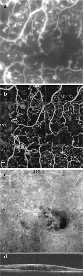
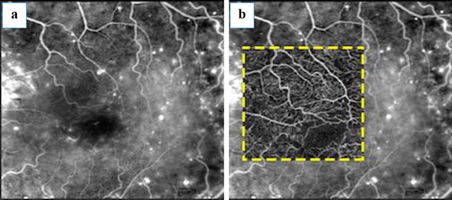
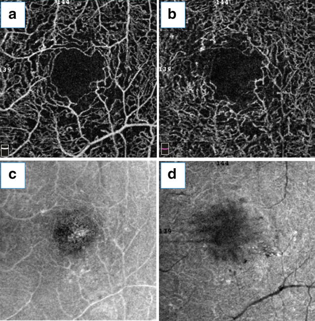
In diabetic retinopathy (DR), macular involvement can present as either macular edema or ischemia. Fluorescein angiography remains the gold standard in the evaluation of retinal vascular perfusion and diagnosis of macular ischemia. However, it is a costly, time-consuming technique, it requires venipuncture, and reports of anaphylaxis and death related to fluorescein injections have been documented, despite their rarity. Optical coherence tomography (OCT) provides a fast and non-invasive method to assess retinal structures at a microscopic level. OCT angiography permits the noninvasive study of retinal and choroid circulation via motion contrast imaging. Split-spectrum amplitude decorrelation angiography combined with OCT angiography has furthered the understanding of retinal and choroidal vascular diseases, allowing the evaluation of retinal microvasculature and identification of subsequent disorders, including DR. Previous studies using OCT angiography have demonstrated that it may demonstrate DR findings such as microaneurysms, arteriolar wall staining, retinal neovascularization, and intraretinal microvascular abnormalities. The purpose of this article is to describe and discuss different concepts regarding OCT angiography, as well as its role in the diagnosis of DR and maculopathy.
在糖尿病性视网膜病变(DR)中,黄斑受累可表现为黄斑水肿或缺血。荧光素血管造影仍是评估视网膜血管灌注和诊断黄斑缺血的金标准。然而,这是一种昂贵、耗时的技术,需要静脉穿刺,并且尽管荧光素注射相关的过敏反应和死亡报告罕见,但已有记录。光学相干断层扫描(OCT)提供了一种快速且非侵入性的方法,可在微观层面评估视网膜结构。OCT血管造影术允许通过运动对比成像对视网膜和脉络膜循环进行非侵入性研究。分裂光谱幅度去相关血管造影术与OCT血管造影术相结合,进一步加深了对视网膜和脉络膜血管疾病的理解,能够评估视网膜微血管系统并识别包括DR在内的后续病症。先前使用OCT血管造影术的研究表明,它可能显示出DR的表现,如微动脉瘤、小动脉壁染色、视网膜新生血管形成和视网膜内微血管异常。本文的目的是描述和讨论关于OCT血管造影术的不同概念,以及它在DR和黄斑病变诊断中的作用。