Ding Ning, Zhao Kui, Lan Yungang, Li Zi, Lv Xiaoling, Su Jingjing, Lu Huijun, Gao Feng, He Wenqi
Key Laboratory of Zoonosis, Ministry of Education, College of Veterinary Medicine, Jilin University Changchun, China.
Key Laboratory of Zoonosis, Ministry of Education, Institute of Zoonosis, Jilin University Changchun, China.
Front Cell Infect Microbiol. 2017 Feb 28;7:56. doi: 10.3389/fcimb.2017.00056. eCollection 2017.
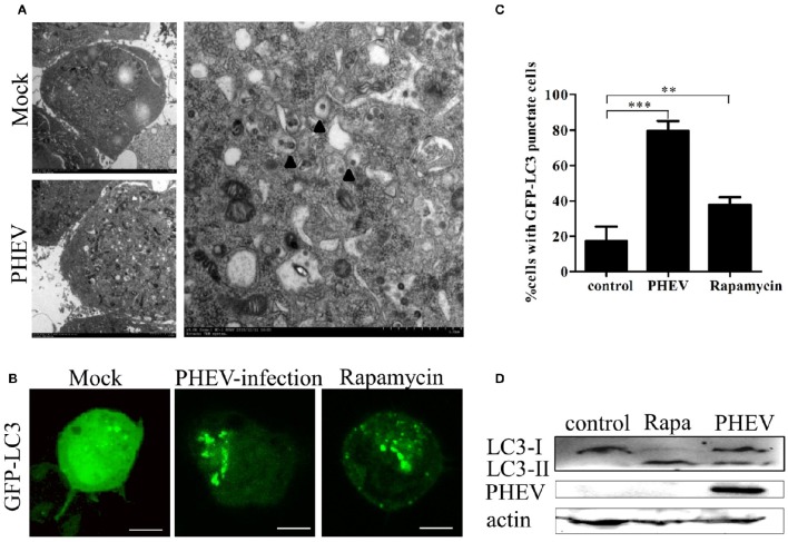
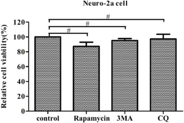
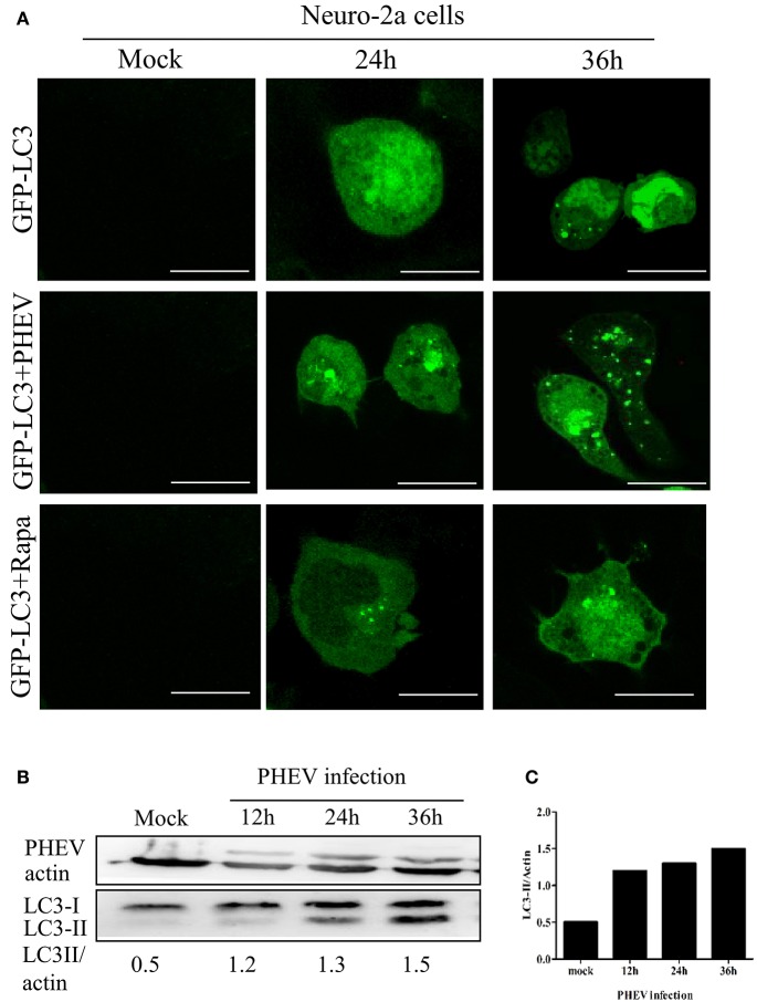
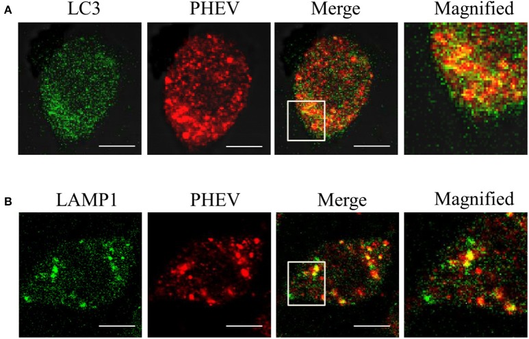
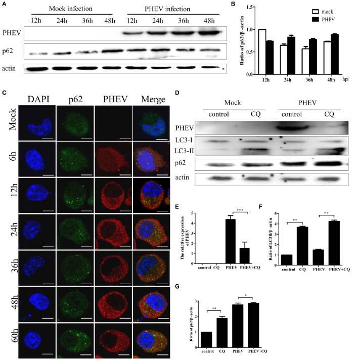
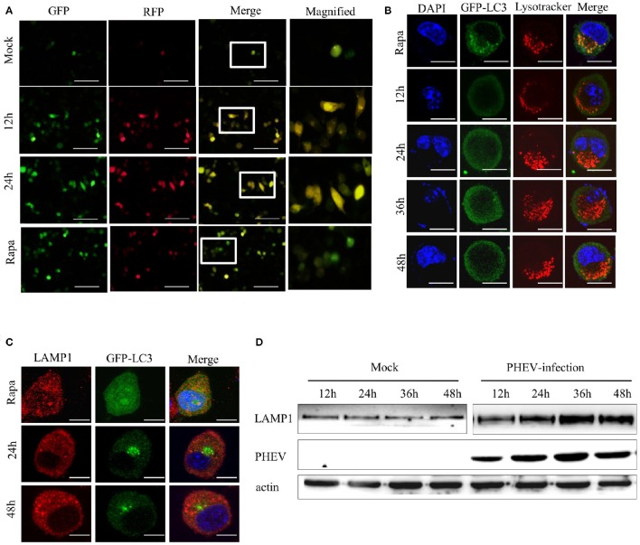
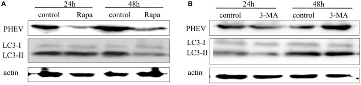
Autophagy is a basic biological metabolic process involving in intracellular membrane transport pathways that recycle cellular components and eliminate intracellular microorganisms within the lysosome. Autophagy also plays an important part in virus infection and propagation. However, some pathogens, including viruses, have evolved unique trick to escape or exploit autophagy. This study explores the mechanism of autophagy induction by porcine hemagglutinating encephalomyelitis virus (PHEV) in Neuro-2a cells, and examines the role of autophagy in PHEV replication. PHEV triggered autophagy in Neuro-2a cells is dependent on the presence of bulk double- or single-membrane vacuoles, the accumulation of GFP-LC3 fluorescent dots, and the LC3 lipidation. In addition, PHEV induced an incomplete autophagic effect because the degradation level of p62 did not change in PHEV-infected cells. Further validation was captured using LysoTracker and lysosome-associated membrane protein by indirect immunofluorescence labeling in PHEV-infected cells. We also investigated the change in viral replication by pharmacological experiments with the autophagy inducer rapamycin or the autophagy inhibitor 3-MA, and the lysosomal inhibitor chloroquine (CQ). Suppression of autophagy by 3-MA increased viral replication, compared with the mock treatment, while promoting of autophagy by rapamycin reduced PHEV replication. CQ treatment enhanced the LC3 lipidation in PHEV-infected Neuro-2a cells but lowered PHEV replication. These results show that PHEV infection induces atypical autophagy and causes the appearance of autophagosomes but blocks the fusion with lysosomes, which is necessary for the replication of PHEV in nerve cells.
自噬是一种基本的生物代谢过程,涉及细胞内膜运输途径,该途径可循环利用细胞成分并在溶酶体内清除细胞内微生物。自噬在病毒感染和传播中也起着重要作用。然而,一些病原体,包括病毒,已经进化出独特的策略来逃避或利用自噬。本研究探讨了猪血凝性脑脊髓炎病毒(PHEV)在Neuro-2a细胞中诱导自噬的机制,并研究了自噬在PHEV复制中的作用。PHEV在Neuro-2a细胞中触发的自噬依赖于大量双膜或单膜空泡的存在、GFP-LC3荧光点的积累以及LC3脂化。此外,PHEV诱导了不完全的自噬效应,因为在PHEV感染的细胞中p62的降解水平没有变化。通过在PHEV感染的细胞中进行间接免疫荧光标记,使用溶酶体示踪剂和溶酶体相关膜蛋白进行了进一步验证。我们还通过用自噬诱导剂雷帕霉素或自噬抑制剂3-MA以及溶酶体抑制剂氯喹(CQ)进行药理学实验,研究了病毒复制的变化。与模拟处理相比,3-MA抑制自噬增加了病毒复制,而雷帕霉素促进自噬则降低了PHEV复制。CQ处理增强了PHEV感染的Neuro-2a细胞中的LC3脂化,但降低了PHEV复制。这些结果表明,PHEV感染诱导非典型自噬并导致自噬体的出现,但阻断了与溶酶体的融合,这是PHEV在神经细胞中复制所必需的。