• 文献检索
  • 文档翻译
  • 深度研究
  • 学术资讯
  • Suppr Zotero 插件Zotero 插件
  • 邀请有礼
  • 套餐&价格
  • 历史记录
应用&插件
Suppr Zotero 插件Zotero 插件浏览器插件Mac 客户端Windows 客户端微信小程序
定价
高级版会员购买积分包购买API积分包
服务
文献检索文档翻译深度研究API 文档MCP 服务
关于我们
关于 Suppr公司介绍联系我们用户协议隐私条款
关注我们

Suppr 超能文献

核心技术专利:CN118964589B侵权必究
粤ICP备2023148730 号-1Suppr @ 2026

文献检索

告别复杂PubMed语法,用中文像聊天一样搜索,搜遍4000万医学文献。AI智能推荐,让科研检索更轻松。

立即免费搜索

文件翻译

保留排版,准确专业,支持PDF/Word/PPT等文件格式,支持 12+语言互译。

免费翻译文档

深度研究

AI帮你快速写综述,25分钟生成高质量综述,智能提取关键信息,辅助科研写作。

立即免费体验

双荧光素与吲哚菁绿血管造影对非流行地区疑似结核性脉络膜视网膜炎评估的贡献

Contribution of Dual Fluorescein and Indocyanine Green Angiography to the Appraisal of Presumed Tuberculous Chorioretinitis in a Non-endemic Area.

作者信息

Massy Raphael, Herbort Carl P

机构信息

Department of Ophthalmology, University of Lausanne, Lausanne, Switzerland; Retinal and Inflammatory Eye Diseases, Centre for Ophthalmic Specialized Care (COS), Teaching Centre Clinic Montchoisi, Lausanne, Switzerland.

出版信息

J Ophthalmic Vis Res. 2017 Jan-Mar;12(1):30-38. doi: 10.4103/2008-322X.200157.

DOI:10.4103/2008-322X.200157
PMID:28299004
原文链接:https://pmc.ncbi.nlm.nih.gov/articles/PMC5340061/
Abstract

PURPOSE

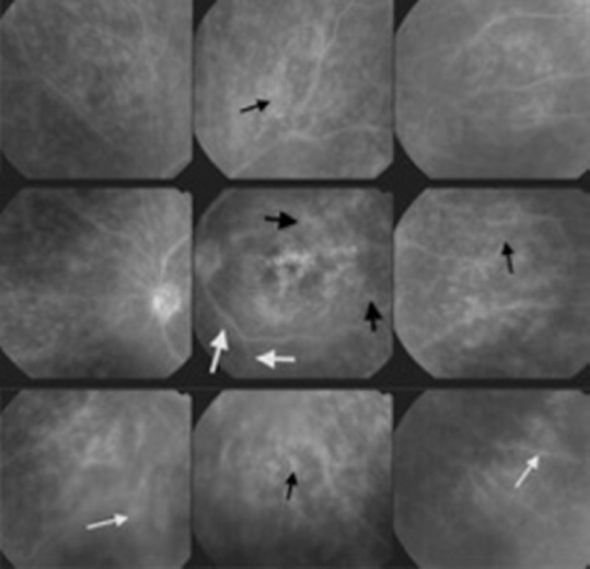
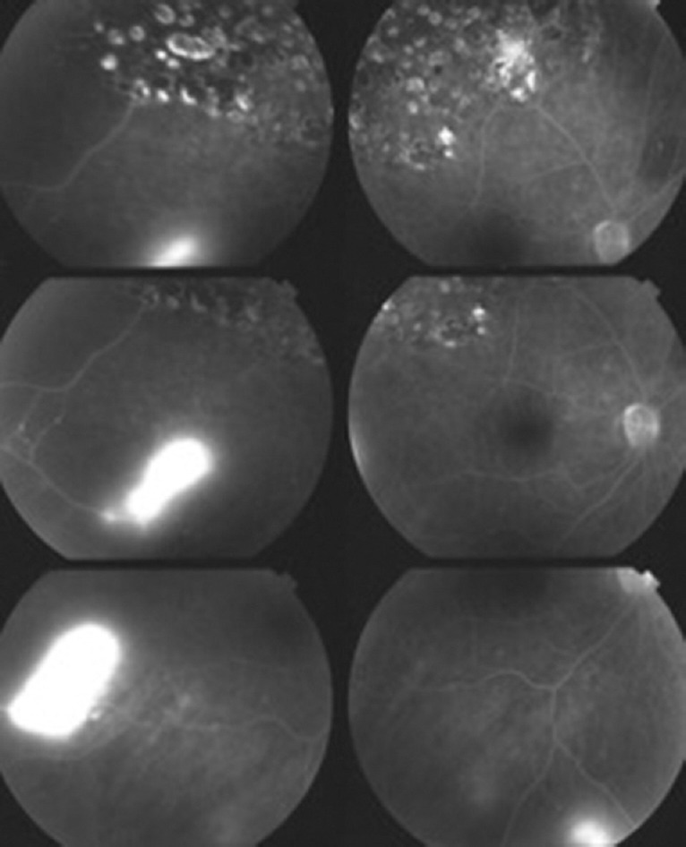
To assess the respective involvement of retina versus choroid in presumed ocular tuberculosis (POT) in a non-endemic area using dual fluorescein (FA) and indocyanine green angiography (ICGA).

METHODS

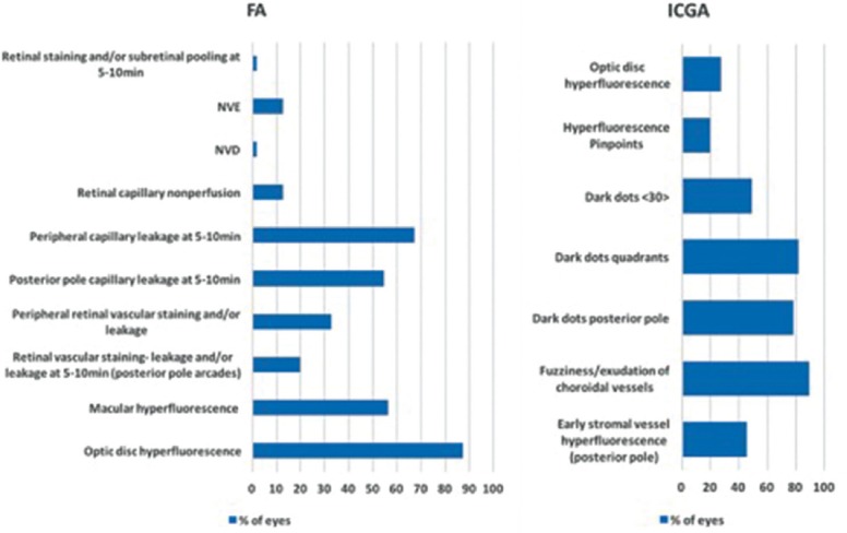
We retrospectively analyzed cases diagnosed with POT at the Centre for Ophthalmic Specialized Care, Lausanne, Switzerland. Angiography signs were quantified using an established dual FA and ICGA scoring system for uveitis.

RESULTS

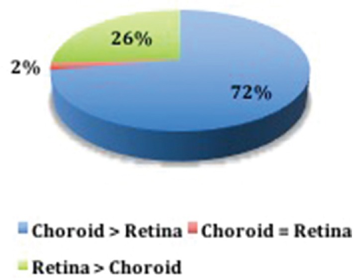
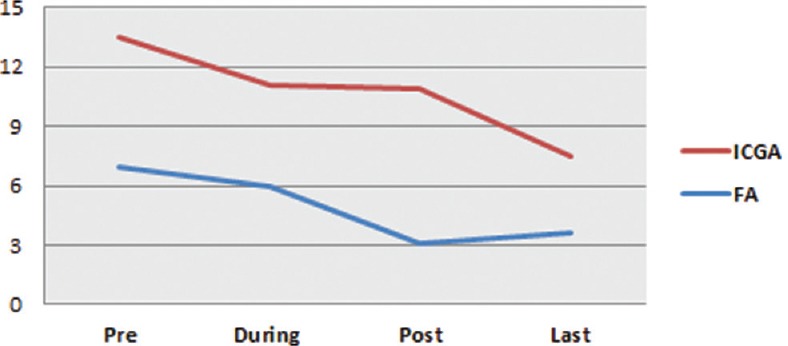
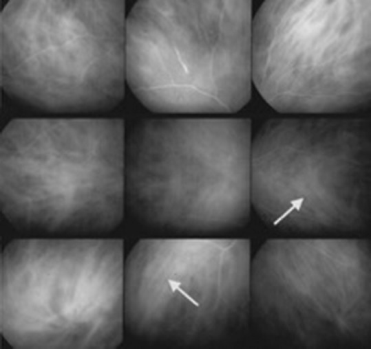
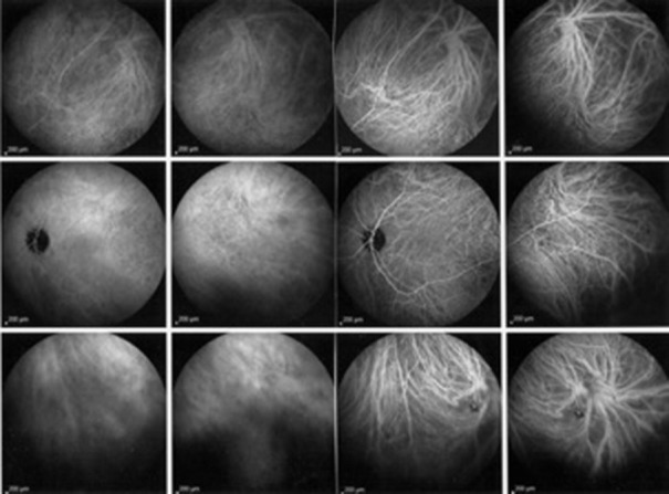
Out of 1739 uveitis patients visited from 1995 to 2014, 53 (3%) were diagnosed with POT; of whom 28 patients (54 eyes) had sufficient data available to be included in this study. Of 54 affected eyes, 39 showed predominant choroidal involvement, 14 showed predominant retinal involvement and one had equal retinal and choroidal scores. Mean angiographic score was 6.97 ± 5.08 for the retina versus 13.48 ± 7.06 for the choroid ( < 0.0001). For patients with sufficient angiographic follow-up after combined anti-tuberculous and inflammation suppressive therapy, mean FA and ICGA scores decreased from 6.97 ± 5.08 to 3.63 ± 3.14 ( = 0.004), and 13.48 ± 7.06 to 7.47 ± 5.58 ( < 0.0001), respectively.

CONCLUSION

These results represent the first report of the respective contributions of retinal and choroidal involvement in POT. Choroidal involvement was more common, for which ICGA is the preferred examination. In cases of compatible uveitis with positive results of an interferon-gamma release assay, particularly in a region that is non-endemic for TB, dual FA and ICGA should be performed to help establish the diagnosis of ocular tuberculosis and improve follow-up.

摘要

目的

在非结核病流行地区,使用双荧光素血管造影(FA)和吲哚菁绿血管造影(ICGA)评估视网膜与脉络膜在疑似眼结核(POT)中的各自受累情况。

方法

我们回顾性分析了在瑞士洛桑眼科专科医院诊断为POT的病例。使用既定的葡萄膜炎双FA和ICGA评分系统对血管造影征象进行量化。

结果

在1995年至2014年就诊的1739例葡萄膜炎患者中,53例(3%)被诊断为POT;其中28例患者(54只眼)有足够的数据可纳入本研究。在54只受累眼中,39只显示主要为脉络膜受累,14只显示主要为视网膜受累,1只视网膜和脉络膜评分相同。视网膜的平均血管造影评分为6.97±5.08,而脉络膜为13.48±7.06(P<0.0001)。对于联合抗结核和抑制炎症治疗后有足够血管造影随访的患者,平均FA和ICGA评分分别从6.97±5.08降至3.63±3.14(P=0.004),从13.48±7.06降至7.47±5.58(P<0.0001)。

结论

这些结果代表了视网膜和脉络膜受累在POT中各自作用的首次报告。脉络膜受累更常见,ICGA是首选检查方法。在葡萄膜炎且干扰素-γ释放试验结果为阳性的情况下,特别是在非结核病流行地区,应进行双FA和ICGA检查以帮助确立眼结核诊断并改善随访。

https://cdn.ncbi.nlm.nih.gov/pmc/blobs/eaa7/5340061/853b01c926d3/JOVR-12-30-g010.jpg
https://cdn.ncbi.nlm.nih.gov/pmc/blobs/eaa7/5340061/ee4ce8fc68d6/JOVR-12-30-g001.jpg
https://cdn.ncbi.nlm.nih.gov/pmc/blobs/eaa7/5340061/96887782717b/JOVR-12-30-g002.jpg
https://cdn.ncbi.nlm.nih.gov/pmc/blobs/eaa7/5340061/bce07dcd3df4/JOVR-12-30-g003.jpg
https://cdn.ncbi.nlm.nih.gov/pmc/blobs/eaa7/5340061/ee4caaf89f4d/JOVR-12-30-g004.jpg
https://cdn.ncbi.nlm.nih.gov/pmc/blobs/eaa7/5340061/1f2973975917/JOVR-12-30-g005.jpg
https://cdn.ncbi.nlm.nih.gov/pmc/blobs/eaa7/5340061/ff92dc90e4ef/JOVR-12-30-g006.jpg
https://cdn.ncbi.nlm.nih.gov/pmc/blobs/eaa7/5340061/12d72870997b/JOVR-12-30-g007.jpg
https://cdn.ncbi.nlm.nih.gov/pmc/blobs/eaa7/5340061/d5fea7308d24/JOVR-12-30-g008.jpg
https://cdn.ncbi.nlm.nih.gov/pmc/blobs/eaa7/5340061/853b01c926d3/JOVR-12-30-g010.jpg

相似文献

1
Contribution of Dual Fluorescein and Indocyanine Green Angiography to the Appraisal of Presumed Tuberculous Chorioretinitis in a Non-endemic Area.双荧光素与吲哚菁绿血管造影对非流行地区疑似结核性脉络膜视网膜炎评估的贡献
J Ophthalmic Vis Res. 2017 Jan-Mar;12(1):30-38. doi: 10.4103/2008-322X.200157.
2
Need for Quantitative Measurement Methods for Posterior Uveitis: Comparison of Dual FA/ICGA Angiography, EDI-OCT Choroidal Thickness and SUN Vitreous Haze Evaluation in Stromal Choroiditis.后葡萄膜炎定量测量方法的必要性:基质性脉络膜炎中荧光素血管造影/吲哚菁绿血管造影双造影、增强深度成像光学相干断层扫描脉络膜厚度测量及SUN玻璃体混浊评估的比较
Klin Monbl Augenheilkd. 2018 Apr;235(4):424-435. doi: 10.1055/s-0043-124966. Epub 2018 Apr 18.
3
Contribution of dual fluorescein and indocyanine green angiography to the appraisal of posterior involvement in birdshot retinochoroiditis and Vogt-Koyanagi-Harada disease.双荧光素和吲哚菁绿血管造影在评估鸟枪弹样视网膜脉络膜炎和小柳原田病后部受累情况中的作用。
Int Ophthalmol. 2018 Apr;38(2):527-539. doi: 10.1007/s10792-017-0487-5. Epub 2017 Mar 15.
4
Comparison of Retinal and Choroidal Involvement in Sarcoidosis-related Chorioretinitis Using Fluorescein and Indocyanine Green Angiography.使用荧光素和吲哚菁绿血管造影术比较结节病相关性脉络膜视网膜炎中视网膜和脉络膜受累情况
J Ophthalmic Vis Res. 2018 Oct-Dec;13(4):426-432. doi: 10.4103/jovr.jovr_201_17.
5
Assessment of measurement methods of posterior inflammation in stromal choroiditis: the value of quantitative outcome measures versus the presently qualitatively based paradigm.基质性脉络膜炎后部炎症测量方法的评估:定量结果测量相对于当前基于定性的范例的价值。
Int Ophthalmol. 2019 Jul;39(7):1567-1574. doi: 10.1007/s10792-018-0979-y. Epub 2018 Jun 26.
6
Assessment and classification of choroidal vasculitis in posterior uveitis using indocyanine green angiography.使用吲哚菁绿血管造影术对后葡萄膜炎中的脉络膜血管炎进行评估和分类。
Klin Monbl Augenheilkd. 2002 Apr;219(4):243-9. doi: 10.1055/s-2002-30661.
7
Scoring of dual fluorescein and ICG inflammatory angiographic signs for the grading of posterior segment inflammation (dual fluorescein and ICG angiographic scoring system for uveitis).用于后段炎症分级的双荧光素和吲哚菁绿炎症血管造影征象评分(葡萄膜炎双荧光素和吲哚菁绿血管造影评分系统)
Int Ophthalmol. 2010 Oct;30(5):539-52. doi: 10.1007/s10792-008-9263-x. Epub 2008 Sep 16.
8
Indocyanine green angiographic features in tuberculous chorioretinitis.结核性脉络膜视网膜炎的吲哚青绿血管造影特征
Am J Ophthalmol. 1999 Mar;127(3):350-3. doi: 10.1016/s0002-9394(98)00325-0.
9
Differential inflammatory involvement in retina and choroïd in birdshot chorioretinopathy.鸟枪弹样脉络膜视网膜病变中视网膜和脉络膜的不同炎症累及情况。
Klin Monbl Augenheilkd. 2004 May;221(5):351-6. doi: 10.1055/s-2004-812827.
10
Retinal vasculitis in HLA-A29 birdshot retinochoroiditis, particularities and imaging narrative of an under-estimated and diagnostic component of the disease.HLA - A29 鸟枪弹样视网膜脉络膜炎中的视网膜血管炎,该疾病一个被低估的诊断组成部分的特点及影像学描述
J Ophthalmic Inflamm Infect. 2024 Jul 9;14(1):31. doi: 10.1186/s12348-024-00406-0.

引用本文的文献

1
Choroidal vasculitis as a biomarker of inflammation of the choroid. Indocyanine Green Angiography (ICGA) spearheading for diagnosis and follow-up, an imaging tutorial.脉络膜血管炎作为脉络膜炎症的生物标志物。吲哚菁绿血管造影(ICGA)在诊断和随访中处于领先地位,影像教程。
J Ophthalmic Inflamm Infect. 2024 Dec 4;14(1):63. doi: 10.1186/s12348-024-00442-w.
2
Retinal vasculitis in HLA-A29 birdshot retinochoroiditis, particularities and imaging narrative of an under-estimated and diagnostic component of the disease.HLA - A29 鸟枪弹样视网膜脉络膜炎中的视网膜血管炎,该疾病一个被低估的诊断组成部分的特点及影像学描述
J Ophthalmic Inflamm Infect. 2024 Jul 9;14(1):31. doi: 10.1186/s12348-024-00406-0.
3

本文引用的文献

1
Diagnosis and treatment for ocular tuberculosis among uveitis specialists: the international perspective.葡萄膜炎专家眼中的眼结核诊断与治疗:国际视角
Ocul Immunol Inflamm. 2015 Feb;23(1):32-9. doi: 10.3109/09273948.2014.994784.
2
Clinics of ocular tuberculosis.眼部结核病诊所
Ocul Immunol Inflamm. 2015 Feb;23(1):14-24. doi: 10.3109/09273948.2014.986582.
3
The usefulness of interferon-gamma release assay for diagnosis of tuberculosis-related uveitis in Korea.γ-干扰素释放试验在韩国诊断结核相关性葡萄膜炎中的应用价值。
Posterior segment inflammatory outcomes assessed using fluorescein angiography in the STOP-UVEITIS study.
在STOP-UVEITIS研究中,使用荧光素血管造影术评估的后段炎症结果。
Int J Retina Vitreous. 2020 Oct 6;6:47. doi: 10.1186/s40942-020-00245-w. eCollection 2020.
4
Advances and potential new developments in imaging techniques for posterior uveitis Part 2: invasive imaging methods.后部葡萄膜炎影像学技术的进展和潜在新发展 第 2 部分:有创性影像学方法。
Eye (Lond). 2021 Jan;35(1):52-73. doi: 10.1038/s41433-020-1072-0. Epub 2020 Aug 10.
5
Comparison of Retinal and Choroidal Involvement in Sarcoidosis-related Chorioretinitis Using Fluorescein and Indocyanine Green Angiography.使用荧光素和吲哚菁绿血管造影术比较结节病相关性脉络膜视网膜炎中视网膜和脉络膜受累情况
J Ophthalmic Vis Res. 2018 Oct-Dec;13(4):426-432. doi: 10.4103/jovr.jovr_201_17.
Korean J Ophthalmol. 2014 Jun;28(3):226-33. doi: 10.3341/kjo.2014.28.3.226. Epub 2014 May 19.
4
Clinical manifestations of patients with intraocular inflammation and positive QuantiFERON-TB gold in-tube test in a country nonendemic for tuberculosis.非结核流行国家眼内炎症患者和阳性 QuantiFERON-TB gold in-tube 试验的临床表现。
Am J Ophthalmol. 2014 Apr;157(4):754-61. doi: 10.1016/j.ajo.2013.11.013. Epub 2013 Nov 18.
5
Interferon γ release assay for the diagnosis of uveitis associated with tuberculosis: a Bayesian evaluation in the absence of a gold standard.结核相关性葡萄膜炎的干扰素 γ 释放试验诊断:无金标准情况下的贝叶斯评价。
Br J Ophthalmol. 2013 Aug;97(8):1062-7. doi: 10.1136/bjophthalmol-2012-302199. Epub 2013 May 30.
6
Diagnosis and treatment of tuberculous uveitis in a low endemic setting.低流行地区结核性葡萄膜炎的诊断与治疗。
Int J Infect Dis. 2013 Nov;17(11):e993-9. doi: 10.1016/j.ijid.2013.03.019. Epub 2013 May 22.
7
Imaging in the diagnosis and management of ocular tuberculosis.
Int Ophthalmol Clin. 2012 Fall;52(4):97-112. doi: 10.1097/IIO.0b013e318265d5a9.
8
Uveitis with occult choroiditis due to Mycobacterium kansasii: limitations of interferon-gamma release assay (IGRA) tests (case report and mini-review on ocular non-tuberculous mycobacteria and IGRA cross-reactivity).堪萨斯分枝杆菌所致伴有隐匿性脉络膜炎的葡萄膜炎:干扰素-γ释放试验(IGRA)检测的局限性(病例报告及关于眼部非结核分枝杆菌和IGRA交叉反应性的小型综述)
Int Ophthalmol. 2012 Oct;32(5):499-506. doi: 10.1007/s10792-012-9588-3. Epub 2012 Jun 2.
9
[Extrapulmonary tuberculosis].[肺外结核病]
Rev Mal Respir. 2012 Apr;29(4):566-78. doi: 10.1016/j.rmr.2011.05.021. Epub 2012 Mar 29.
10
Tuberculosis-related choriocapillaritis (multifocal-serpiginous choroiditis): follow-up and precise monitoring of therapy by indocyanine green angiography.结核相关性脉络膜毛细血管炎(多灶性匐行性脉络膜炎):通过吲哚菁绿血管造影术进行随访及精确的治疗监测
Int Ophthalmol. 2012 Feb;32(1):55-60. doi: 10.1007/s10792-011-9508-y. Epub 2012 Jan 17.