• 文献检索
  • 文档翻译
  • 深度研究
  • 学术资讯
  • Suppr Zotero 插件Zotero 插件
  • 邀请有礼
  • 套餐&价格
  • 历史记录
应用&插件
Suppr Zotero 插件Zotero 插件浏览器插件Mac 客户端Windows 客户端微信小程序
定价
高级版会员购买积分包购买API积分包
服务
文献检索文档翻译深度研究API 文档MCP 服务
关于我们
关于 Suppr公司介绍联系我们用户协议隐私条款
关注我们

Suppr 超能文献

核心技术专利:CN118964589B侵权必究
粤ICP备2023148730 号-1Suppr @ 2026

文献检索

告别复杂PubMed语法,用中文像聊天一样搜索,搜遍4000万医学文献。AI智能推荐,让科研检索更轻松。

立即免费搜索

文件翻译

保留排版,准确专业,支持PDF/Word/PPT等文件格式,支持 12+语言互译。

免费翻译文档

深度研究

AI帮你快速写综述,25分钟生成高质量综述,智能提取关键信息,辅助科研写作。

立即免费体验

使用荧光素和吲哚菁绿血管造影术比较结节病相关性脉络膜视网膜炎中视网膜和脉络膜受累情况

Comparison of Retinal and Choroidal Involvement in Sarcoidosis-related Chorioretinitis Using Fluorescein and Indocyanine Green Angiography.

作者信息

El Ameen Ala'a, Herbort Carl P

机构信息

Retinal and Inflammatory Eye Diseases, Centre for Ophthalmic Specialised Care, Teaching Centre Clinic Montchoisi, Lausanne, Switzerland.

Department of Ophthalmology, University of Lausanne, Lausanne, Switzerland.

出版信息

J Ophthalmic Vis Res. 2018 Oct-Dec;13(4):426-432. doi: 10.4103/jovr.jovr_201_17.

DOI:10.4103/jovr.jovr_201_17
PMID:30479712
原文链接:https://pmc.ncbi.nlm.nih.gov/articles/PMC6210872/
Abstract

PURPOSE

To compare the involvement of the retina with that of the choroid in ocular sarcoidosis (OS) using dual fluorescein angiography (FA)/indocyanine green angiography (ICGA).

METHODS

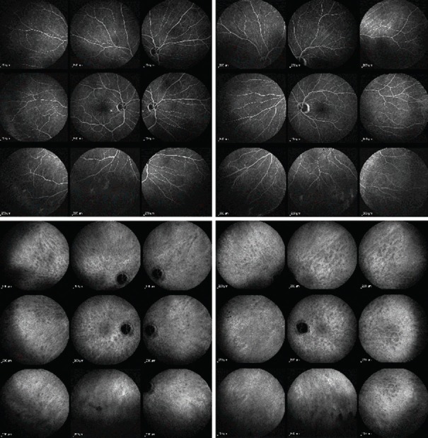
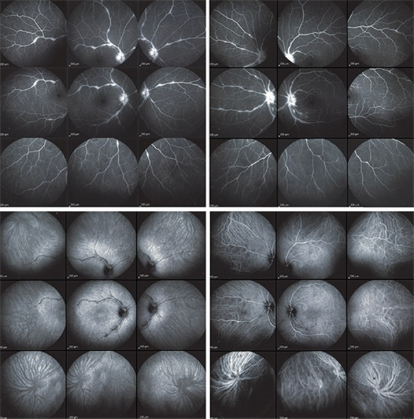
A retrospective study of 23 patients with the diagnosis of OS was performed. Angiographic signs were quantified following the established FA/ICGA scoring system for uveitis.

RESULTS

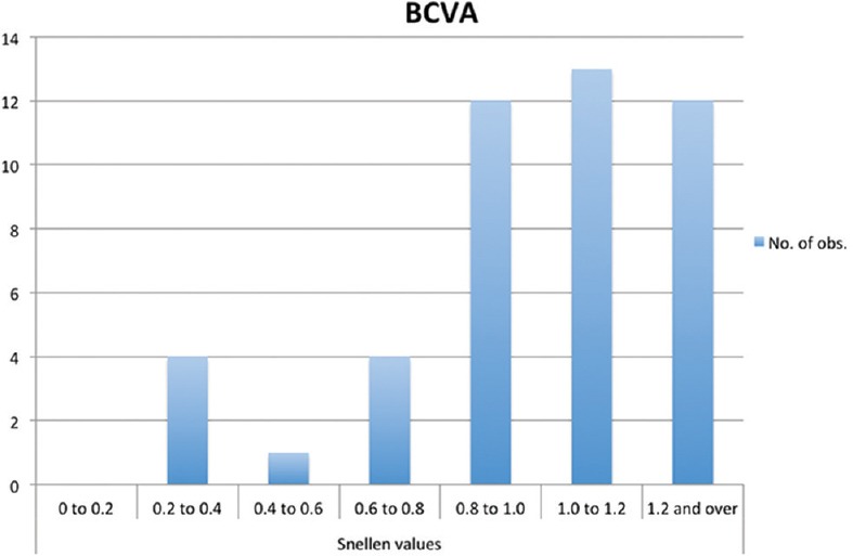
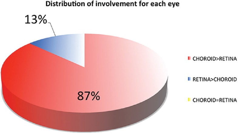
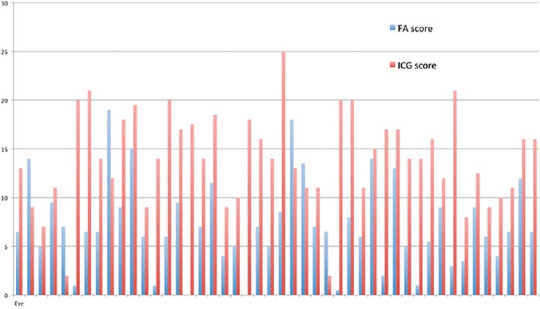
The choroid was predominantly involved in 19 (82.6%) patients or 87% (40/46) of the eyes, and the retina in 2 (8.7%) patients or 13% (6/46) of the eyes. The mean angiographic score was 7.15 ± 4.5 for the retina (FA) compared to 14.02 ± 4.86 for the choroid (ICGA) ( < 0.0001). In 13% (3/23) of patients, FA did not show retinal inflammation, whereas ICGA was strongly positive, revealing occult choroidal lesions.

CONCLUSION

The choroid is preferentially involved in OS, for which ICGA is the examination of choice. There is a risk of underestimating the global ocular involvement and of missing choroidal involvement if only FA is used. FA/ICGA scoring system allows for quantitative assessment of inflammation in the posterior uvea that occurs in OS; therefore, the system can be useful to quantitatively monitor outcomes in clinical trials.

摘要

目的

使用双荧光素血管造影(FA)/吲哚菁绿血管造影(ICGA)比较眼部结节病(OS)中视网膜与脉络膜受累情况。

方法

对23例诊断为OS的患者进行回顾性研究。按照既定的葡萄膜炎FA/ICGA评分系统对血管造影征象进行量化。

结果

脉络膜受累为主的患者有19例(82.6%),占所有眼睛的87%(40/46);视网膜受累为主的患者有2例(8.7%),占所有眼睛的13%(6/46)。视网膜(FA)的平均血管造影评分为7.15±4.5,而脉络膜(ICGA)为14.02±4.86(P<0.0001)。13%(3/23)的患者FA未显示视网膜炎症,而ICGA呈强阳性,揭示了隐匿性脉络膜病变。

结论

脉络膜在OS中更易受累,ICGA是首选检查方法。若仅使用FA,存在低估整体眼部受累情况及漏诊脉络膜受累的风险。FA/ICGA评分系统可对OS中后葡萄膜的炎症进行定量评估;因此,该系统有助于在临床试验中对结果进行定量监测。

https://cdn.ncbi.nlm.nih.gov/pmc/blobs/0be3/6210872/c1b6c7d029ee/JOVR-13-426-g007.jpg
https://cdn.ncbi.nlm.nih.gov/pmc/blobs/0be3/6210872/50ce08f5726a/JOVR-13-426-g001.jpg
https://cdn.ncbi.nlm.nih.gov/pmc/blobs/0be3/6210872/fe4c33c49b79/JOVR-13-426-g002.jpg
https://cdn.ncbi.nlm.nih.gov/pmc/blobs/0be3/6210872/16243d50b6d3/JOVR-13-426-g003.jpg
https://cdn.ncbi.nlm.nih.gov/pmc/blobs/0be3/6210872/9341a877b363/JOVR-13-426-g004.jpg
https://cdn.ncbi.nlm.nih.gov/pmc/blobs/0be3/6210872/379ae065cb05/JOVR-13-426-g005.jpg
https://cdn.ncbi.nlm.nih.gov/pmc/blobs/0be3/6210872/ae71d6a658e2/JOVR-13-426-g006.jpg
https://cdn.ncbi.nlm.nih.gov/pmc/blobs/0be3/6210872/c1b6c7d029ee/JOVR-13-426-g007.jpg

相似文献

1
Comparison of Retinal and Choroidal Involvement in Sarcoidosis-related Chorioretinitis Using Fluorescein and Indocyanine Green Angiography.使用荧光素和吲哚菁绿血管造影术比较结节病相关性脉络膜视网膜炎中视网膜和脉络膜受累情况
J Ophthalmic Vis Res. 2018 Oct-Dec;13(4):426-432. doi: 10.4103/jovr.jovr_201_17.
2
Need for Quantitative Measurement Methods for Posterior Uveitis: Comparison of Dual FA/ICGA Angiography, EDI-OCT Choroidal Thickness and SUN Vitreous Haze Evaluation in Stromal Choroiditis.后葡萄膜炎定量测量方法的必要性:基质性脉络膜炎中荧光素血管造影/吲哚菁绿血管造影双造影、增强深度成像光学相干断层扫描脉络膜厚度测量及SUN玻璃体混浊评估的比较
Klin Monbl Augenheilkd. 2018 Apr;235(4):424-435. doi: 10.1055/s-0043-124966. Epub 2018 Apr 18.
3
Contribution of Dual Fluorescein and Indocyanine Green Angiography to the Appraisal of Presumed Tuberculous Chorioretinitis in a Non-endemic Area.双荧光素与吲哚菁绿血管造影对非流行地区疑似结核性脉络膜视网膜炎评估的贡献
J Ophthalmic Vis Res. 2017 Jan-Mar;12(1):30-38. doi: 10.4103/2008-322X.200157.
4
Contribution of dual fluorescein and indocyanine green angiography to the appraisal of posterior involvement in birdshot retinochoroiditis and Vogt-Koyanagi-Harada disease.双荧光素和吲哚菁绿血管造影在评估鸟枪弹样视网膜脉络膜炎和小柳原田病后部受累情况中的作用。
Int Ophthalmol. 2018 Apr;38(2):527-539. doi: 10.1007/s10792-017-0487-5. Epub 2017 Mar 15.
5
Assessment of measurement methods of posterior inflammation in stromal choroiditis: the value of quantitative outcome measures versus the presently qualitatively based paradigm.基质性脉络膜炎后部炎症测量方法的评估:定量结果测量相对于当前基于定性的范例的价值。
Int Ophthalmol. 2019 Jul;39(7):1567-1574. doi: 10.1007/s10792-018-0979-y. Epub 2018 Jun 26.
6
Assessment and classification of choroidal vasculitis in posterior uveitis using indocyanine green angiography.使用吲哚菁绿血管造影术对后葡萄膜炎中的脉络膜血管炎进行评估和分类。
Klin Monbl Augenheilkd. 2002 Apr;219(4):243-9. doi: 10.1055/s-2002-30661.
7
Scoring of dual fluorescein and ICG inflammatory angiographic signs for the grading of posterior segment inflammation (dual fluorescein and ICG angiographic scoring system for uveitis).用于后段炎症分级的双荧光素和吲哚菁绿炎症血管造影征象评分(葡萄膜炎双荧光素和吲哚菁绿血管造影评分系统)
Int Ophthalmol. 2010 Oct;30(5):539-52. doi: 10.1007/s10792-008-9263-x. Epub 2008 Sep 16.
8
Fluorescein and indocyanine green angiography findings in Behçet's disease.白塞病的荧光素和吲哚菁绿血管造影结果。
Br J Ophthalmol. 2003 Dec;87(12):1466-8. doi: 10.1136/bjo.87.12.1466.
9
Inflammation of the posterior uvea: findings on fundus fluorescein and indocyanine green angiography.后葡萄膜炎:眼底荧光素和吲哚菁绿血管造影检查结果
Ocul Immunol Inflamm. 2006 Jun;14(3):171-9. doi: 10.1080/09273940600660524.
10
Indocyanine green angiographic findings in serpiginous choroidopathy.匐行性脉络膜病变的吲哚菁绿血管造影表现
Br J Ophthalmol. 1996 Jun;80(6):536-40. doi: 10.1136/bjo.80.6.536.

引用本文的文献

1
Inadequacies in uveitis: misnomers, incongruencies, persistence of obsolete terminologies & inappropriate guidelines, treatment inadequacies, and misinterpretations.葡萄膜炎的不足之处:名称不当、不一致、过时术语的持续存在及不恰当的指南、治疗不足和误解。
J Ophthalmic Inflamm Infect. 2025 Mar 6;15(1):19. doi: 10.1186/s12348-025-00470-0.
2
Choroidal vasculitis as a biomarker of inflammation of the choroid. Indocyanine Green Angiography (ICGA) spearheading for diagnosis and follow-up, an imaging tutorial.脉络膜血管炎作为脉络膜炎症的生物标志物。吲哚菁绿血管造影(ICGA)在诊断和随访中处于领先地位,影像教程。
J Ophthalmic Inflamm Infect. 2024 Dec 4;14(1):63. doi: 10.1186/s12348-024-00442-w.
3

本文引用的文献

1
Contribution of Dual Fluorescein and Indocyanine Green Angiography to the Appraisal of Presumed Tuberculous Chorioretinitis in a Non-endemic Area.双荧光素与吲哚菁绿血管造影对非流行地区疑似结核性脉络膜视网膜炎评估的贡献
J Ophthalmic Vis Res. 2017 Jan-Mar;12(1):30-38. doi: 10.4103/2008-322X.200157.
2
QUANTITATIVE ANALYSIS OF STRUCTURAL ALTERATIONS IN THE CHOROID OF PATIENTS WITH ACTIVE BEHÇET UVEITIS.活动性 Behcet 葡萄膜炎患者脉络膜结构改变的定量分析。
Retina. 2018 Apr;38(4):828-840. doi: 10.1097/IAE.0000000000001587.
3
Failure to Integrate Quantitative Measurement Methods of Ocular Inflammation Hampers Clinical Practice and Trials on New Therapies for Posterior Uveitis.
Retinal vasculitis in HLA-A29 birdshot retinochoroiditis, particularities and imaging narrative of an under-estimated and diagnostic component of the disease.
HLA - A29 鸟枪弹样视网膜脉络膜炎中的视网膜血管炎,该疾病一个被低估的诊断组成部分的特点及影像学描述
J Ophthalmic Inflamm Infect. 2024 Jul 9;14(1):31. doi: 10.1186/s12348-024-00406-0.
4
Optical Coherence Tomography Angiography (OCT-A) in Uveitis: A Literature Review and a Reassessment of Its Real Role.葡萄膜炎中的光学相干断层扫描血管造影(OCT-A):文献综述及其实际作用的重新评估
Diagnostics (Basel). 2023 Feb 7;13(4):601. doi: 10.3390/diagnostics13040601.
5
Benefits and Limitations of OCT-A in the Diagnosis and Follow-Up of Posterior Intraocular Inflammation in Current Clinical Practice: A Valuable Tool or a Deceiver?光学相干断层扫描血管造影(OCT-A)在当前临床实践中诊断和随访眼后段炎症的益处与局限性:是有用工具还是误导?
Diagnostics (Basel). 2022 Sep 30;12(10):2384. doi: 10.3390/diagnostics12102384.
6
Sarcoid Uveitis: An Intriguing Challenger.结节性葡萄膜炎:一个引人入胜的挑战。
Medicina (Kaunas). 2022 Jul 4;58(7):898. doi: 10.3390/medicina58070898.
7
Clinicopathology of non-infectious choroiditis: evolution of its appraisal during the last 2-3 decades from "white dot syndromes" to precise classification.非感染性脉络膜炎的临床病理学:过去20至30年间其评估从“白点综合征”到精确分类的演变
J Ophthalmic Inflamm Infect. 2021 Nov 17;11(1):43. doi: 10.1186/s12348-021-00274-y.
8
Precise, simplified diagnostic criteria and optimised management of initial-onset Vogt-Koyanagi-Harada disease: an updated review.初发性 Vogt-小柳-原田病的精确简化诊断标准和优化管理:最新综述。
Eye (Lond). 2022 Jan;36(1):29-43. doi: 10.1038/s41433-021-01573-3. Epub 2021 Jun 18.
9
A Comprehensive Review of Sarcoidosis Diagnosis and Monitoring for the Pulmonologist.肺科医生结节病诊断与监测的全面综述
Pulm Ther. 2021 Dec;7(2):309-324. doi: 10.1007/s41030-021-00161-w. Epub 2021 Jun 6.
10
Classification of Non-Infectious and/or Immune Mediated Choroiditis: A Brief Overview of the Essentials.非感染性和/或免疫介导性脉络膜炎的分类:要点概述
Diagnostics (Basel). 2021 May 24;11(6):939. doi: 10.3390/diagnostics11060939.
未能整合眼部炎症的定量测量方法阻碍了后葡萄膜炎新疗法的临床实践和试验。
J Ocul Pharmacol Ther. 2017 May;33(4):263-277. doi: 10.1089/jop.2016.0089. Epub 2016 Dec 12.
4
Interobserver agreement in scoring of dual fluorescein and ICG inflammatory angiographic signs for the grading of posterior segment inflammation.评估后段炎症时,双荧光素和吲哚菁绿炎性血管造影征象的评分中观察者间一致性。
Ocul Immunol Inflamm. 2010 Oct;18(5):385-9. doi: 10.3109/09273948.2010.489730.
5
International criteria for the diagnosis of ocular sarcoidosis: results of the first International Workshop On Ocular Sarcoidosis (IWOS).眼部结节病的国际诊断标准:首届眼部结节病国际研讨会(IWOS)的结果
Ocul Immunol Inflamm. 2009 May-Jun;17(3):160-9. doi: 10.1080/09273940902818861.
6
Suboptimal therapy controls clinically apparent disease but not subclinical progression of Vogt-Koyanagi-Harada disease.次优治疗可控制伏格特-小柳-原田病的临床显性疾病,但无法控制其亚临床进展。
Int Ophthalmol. 2010 Feb;30(1):41-50. doi: 10.1007/s10792-008-9288-1. Epub 2009 Jan 17.
7
Scoring of dual fluorescein and ICG inflammatory angiographic signs for the grading of posterior segment inflammation (dual fluorescein and ICG angiographic scoring system for uveitis).用于后段炎症分级的双荧光素和吲哚菁绿炎症血管造影征象评分(葡萄膜炎双荧光素和吲哚菁绿血管造影评分系统)
Int Ophthalmol. 2010 Oct;30(5):539-52. doi: 10.1007/s10792-008-9263-x. Epub 2008 Sep 16.
8
Standardization of uveitis nomenclature for reporting clinical data. Results of the First International Workshop.用于报告临床数据的葡萄膜炎命名标准化。第一次国际研讨会的结果。
Am J Ophthalmol. 2005 Sep;140(3):509-16. doi: 10.1016/j.ajo.2005.03.057.
9
Precise monitoring and differentiation of inflammatory events by indocyanine green (ICG) angiography in a case of recurrent posterior sarcoid uveitis.吲哚菁绿(ICG)血管造影术在复发性后部结节病性葡萄膜炎病例中对炎症事件的精确监测与鉴别
Ocul Immunol Inflamm. 2000 Dec;8(4):303-6. doi: 10.1076/ocii.8.4.303.6458.
10
The contribution of indocyanine green angiography to the appraisal and management of Vogt-Koyanagi-Harada disease.吲哚青绿血管造影在Vogt-小柳-原田病评估与治疗中的作用。
Ophthalmology. 2001 Jan;108(1):54-64. doi: 10.1016/s0161-6420(00)00428-0.