Li Hong, Yang Tiandi, Liao Tingting, Debowski Aleksandra W, Nilsson Hans-Olof, Fulurija Alma, Haslam Stuart M, Mulloy Barbara, Dell Anne, Stubbs Keith A, Marshall Barry J, Benghezal Mohammed
West China Marshall Research Center for Infectious Diseases, Center of Infectious Diseases, Division of Infectious Diseases, State Key Laboratory of Biotherapy, West China Hospital, Sichuan University, Chengdu, China.
Helicobacter pylori Research Laboratory and Ondek Pty Ltd., School of Pathology & Laboratory Medicine, Marshall Centre for Infectious Disease Research and Training, University of Western Australia, Nedlands, Australia.
PLoS Pathog. 2017 Mar 17;13(3):e1006280. doi: 10.1371/journal.ppat.1006280. eCollection 2017 Mar.
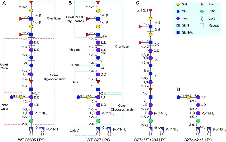
Helicobacter pylori lipopolysaccharide promotes chronic gastric colonisation through O-antigen host mimicry and resistance to mucosal antimicrobial peptides mediated primarily by modifications of the lipid A. The structural organisation of the core and O-antigen domains of H. pylori lipopolysaccharide remains unclear, as the O-antigen attachment site has still to be identified experimentally. Here, structural investigations of lipopolysaccharides purified from two wild-type strains and the O-antigen ligase mutant revealed that the H. pylori core-oligosaccharide domain is a short conserved hexasaccharide (Glc-Gal-DD-Hep-LD-Hep-LD-Hep-KDO) decorated with the O-antigen domain encompassing a conserved trisaccharide (-DD-Hep-Fuc-GlcNAc-) and variable glucan, heptan and Lewis antigens. Furthermore, the putative heptosyltransferase HP1284 was found to be required for the transfer of the third heptose residue to the core-oligosaccharide. Interestingly, mutation of HP1284 did not affect the ligation of the O-antigen and resulted in the attachment of the O-antigen onto an incomplete core-oligosaccharide missing the third heptose and the adjoining Glc-Gal residues. Mutants deficient in either HP1284 or O-antigen ligase displayed a moderate increase in susceptibility to polymyxin B but were unable to colonise the mouse gastric mucosa. Finally, mapping mutagenesis and colonisation data of previous studies onto the redefined organisation of H. pylori lipopolysaccharide revealed that only the conserved motifs were essential for colonisation. In conclusion, H. pylori lipopolysaccharide is missing the canonical inner and outer core organisation. Instead it displays a short core and a longer O-antigen encompassing residues previously assigned as the outer core domain. The redefinition of H. pylori lipopolysaccharide domains warrants future studies to dissect the role of each domain in host-pathogen interactions. Also enzymes involved in the assembly of the conserved core structure, such as HP1284, could be attractive targets for the design of new therapeutic agents for managing persistent H. pylori infection causing peptic ulcers and gastric cancer.
幽门螺杆菌脂多糖通过O抗原模拟宿主以及对主要由脂质A修饰介导的黏膜抗菌肽产生抗性来促进慢性胃定植。幽门螺杆菌脂多糖核心和O抗原结构域的组织结构仍不清楚,因为O抗原连接位点仍有待通过实验确定。在这里,对从两种野生型菌株和O抗原连接酶突变体中纯化的脂多糖进行的结构研究表明,幽门螺杆菌核心寡糖结构域是一种短的保守六糖(葡萄糖-半乳糖-DD-庚糖-LD-庚糖-LD-庚糖-KDO),装饰有O抗原结构域,该结构域包含一个保守的三糖(-DD-庚糖-岩藻糖-氨基葡萄糖-)以及可变的葡聚糖、庚聚糖和Lewis抗原。此外,发现假定的庚糖基转移酶HP1284是将第三个庚糖残基转移到核心寡糖所必需的。有趣的是,HP1284的突变并不影响O抗原的连接,并导致O抗原附着在缺少第三个庚糖以及相邻葡萄糖-半乳糖残基的不完整核心寡糖上。缺乏HP1284或O抗原连接酶的突变体对多粘菌素B的敏感性适度增加,但无法在小鼠胃黏膜中定植。最后,将先前研究的定位诱变和定植数据映射到重新定义的幽门螺杆菌脂多糖组织上,发现只有保守基序对定植至关重要。总之,幽门螺杆菌脂多糖缺少典型的内核心和外核心组织。相反,它显示出一个短的核心和一个更长的O抗原,其中包含以前被指定为外核心结构域的残基。幽门螺杆菌脂多糖结构域的重新定义值得未来开展研究,以剖析每个结构域在宿主-病原体相互作用中的作用。此外参与保守核心结构组装的酶,如HP1284,可能是设计用于治疗由幽门螺杆菌持续感染引起的消化性溃疡和胃癌的新型治疗药物的有吸引力的靶点。