Ajiboye Taofeek O, Habibu Ramat S, Saidu Kabiru, Haliru Fatimah Z, Ajiboye Hikmat O, Aliyu Najeeb O, Ibitoye Oluwayemisi B, Uwazie Judith N, Muritala Hamdalat F, Bello Sharafa A, Yusuf Idris I, Mohammed Aisha O
Antioxidants, Redox Biology and Toxicology Research Laboratory, Department of Biological Sciences, Al-Hikmah University, Ilorin, Nigeria.
Department of Biochemistry, University of Ilorin, Ilorin, Nigeria.
Microbiologyopen. 2017 Aug;6(4). doi: 10.1002/mbo3.472. Epub 2017 Mar 27.
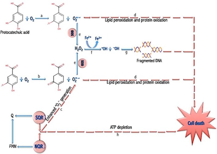
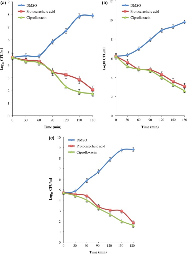
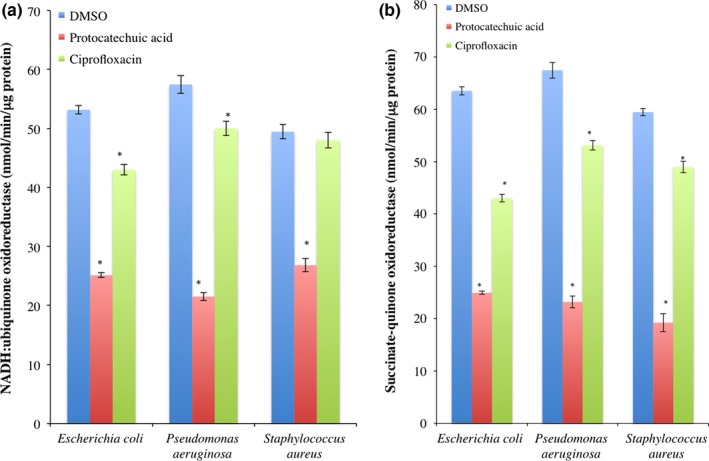
The involvement of oxidative stress in protocatechuic acid-mediated bacterial lethality was investigated. Minimum inhibitory concentrations (MIC) and minimum bactericidal concentration (MBC) of protocatechuic acid against Escherichia coli, Pseudomonas aeruginosa, and Staphylococcus aureus are 600 and 700 μg/ml, 600 and 800 μg/ml, and 600 and 800 μg/ml, respectively. The optical densities and colony-forming units of protocatechuic acid-treated bacteria decreased in time-dependent manner. Protocatechuic acid (4× MIC) significantly increased the superoxide anion content of E. coli, P. aeruginosa, and S. aureus compared to dimethyl sulfoxide (DMSO). Superoxide dismutase, catalase, and NAD /NADH in protocatechuic acid-treated E. coli, P. aeruginosa, and S. aureus increased significantly when compared to DMSO. Conversely, level of reduced glutathione decreased in protocatechuic acid-treated E. coli, P. aeruginosa, and S. aureus, while glutathione disulfide increased when compared to DMSO. Furthermore, malondialdehyde and fragmented DNA increased significantly following exposure to protocatechuic acid. Protocatechuic acid inhibited the activity of complexes I and II. From the above findings, protocatechuic acid enhanced the generation of reactive oxygen species (superoxide anion radical and hydroxyl radical) in E. coli, P. aeruginosa, and S. aureus, possibly by autoxidation, fenton chemistry, and inhibiting electron transport chain resulting in lipid peroxidation and DNA fragmentation and consequentially bacterial cell death.
研究了氧化应激在原儿茶酸介导的细菌致死性中的作用。原儿茶酸对大肠杆菌、铜绿假单胞菌和金黄色葡萄球菌的最低抑菌浓度(MIC)和最低杀菌浓度(MBC)分别为600和700μg/ml、600和800μg/ml、600和800μg/ml。经原儿茶酸处理的细菌的光密度和菌落形成单位呈时间依赖性下降。与二甲基亚砜(DMSO)相比,原儿茶酸(4×MIC)显著增加了大肠杆菌、铜绿假单胞菌和金黄色葡萄球菌的超氧阴离子含量。与DMSO相比,经原儿茶酸处理的大肠杆菌、铜绿假单胞菌和金黄色葡萄球菌中的超氧化物歧化酶、过氧化氢酶和NAD/NADH显著增加。相反,经原儿茶酸处理的大肠杆菌、铜绿假单胞菌和金黄色葡萄球菌中还原型谷胱甘肽水平降低,而与DMSO相比,氧化型谷胱甘肽增加。此外,暴露于原儿茶酸后,丙二醛和片段化DNA显著增加。原儿茶酸抑制复合物I和II的活性。根据上述发现,原儿茶酸可能通过自氧化、芬顿化学反应和抑制电子传递链,增强了大肠杆菌、铜绿假单胞菌和金黄色葡萄球菌中活性氧(超氧阴离子自由基和羟基自由基)的生成,导致脂质过氧化和DNA片段化,进而导致细菌细胞死亡。