• 文献检索
  • 文档翻译
  • 深度研究
  • 学术资讯
  • Suppr Zotero 插件Zotero 插件
  • 邀请有礼
  • 套餐&价格
  • 历史记录
应用&插件
Suppr Zotero 插件Zotero 插件浏览器插件Mac 客户端Windows 客户端微信小程序
定价
高级版会员购买积分包购买API积分包
服务
文献检索文档翻译深度研究API 文档MCP 服务
关于我们
关于 Suppr公司介绍联系我们用户协议隐私条款
关注我们

Suppr 超能文献

核心技术专利:CN118964589B侵权必究
粤ICP备2023148730 号-1Suppr @ 2026

文献检索

告别复杂PubMed语法,用中文像聊天一样搜索,搜遍4000万医学文献。AI智能推荐,让科研检索更轻松。

立即免费搜索

文件翻译

保留排版,准确专业,支持PDF/Word/PPT等文件格式,支持 12+语言互译。

免费翻译文档

深度研究

AI帮你快速写综述,25分钟生成高质量综述,智能提取关键信息,辅助科研写作。

立即免费体验

通过光学相位阈值法进行稳健的微生物细胞分割,处理要求极低。

Robust microbial cell segmentation by optical-phase thresholding with minimal processing requirements.

作者信息

Alanazi H, Canul A J, Garman A, Quimby J, Vasdekis A E

机构信息

Department of Physics, University of Idaho, Moscow, Idaho, 83844.

出版信息

Cytometry A. 2017 May;91(5):443-449. doi: 10.1002/cyto.a.23099. Epub 2017 Mar 30.

DOI:10.1002/cyto.a.23099
PMID:28371011
原文链接:https://pmc.ncbi.nlm.nih.gov/articles/PMC6585648/
Abstract

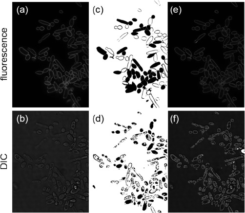
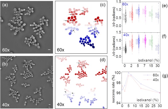
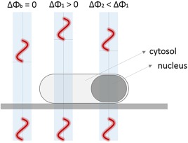
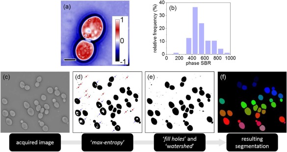
High-throughput imaging with single-cell resolution has enabled remarkable discoveries in cell physiology and Systems Biology investigations. A common, and often the most challenging step in all such imaging implementations, is the ability to segment multiple images to regions that correspond to individual cells. Here, a robust segmentation strategy for microbial cells using Quantitative Phase Imaging is reported. The proposed method enables a greater than 99% yeast cell segmentation success rate, without any computationally-intensive, post-acquisition processing. We also detail how the method can be expanded to bacterial cell segmentation with 98% success rates with substantially reduced processing requirements in comparison to existing methods. We attribute this improved performance to the remarkably uniform background, elimination of cell-to-cell and intracellular optical artifacts, and enhanced signal-to-background ratio-all innate properties of imaging in the optical-phase domain. © 2017 International Society for Advancement of Cytometry.

摘要

具有单细胞分辨率的高通量成像技术在细胞生理学和系统生物学研究中带来了显著发现。在所有此类成像实现中,一个常见且往往最具挑战性的步骤是能够将多个图像分割成对应于单个细胞的区域。在此,报道了一种使用定量相成像技术对微生物细胞进行稳健分割的策略。所提出的方法能够实现超过99%的酵母细胞分割成功率,且无需任何计算密集型的采集后处理。我们还详细说明了该方法如何能够扩展到细菌细胞分割,成功率达到98%,与现有方法相比,处理要求大幅降低。我们将这种性能提升归因于光学相域成像固有的显著均匀背景、细胞间和细胞内光学伪像的消除以及增强的信噪比。© 2017国际细胞计量学促进协会。

https://cdn.ncbi.nlm.nih.gov/pmc/blobs/d6f7/6585648/17a1d923a6e8/CYTO-91-443-g004.jpg
https://cdn.ncbi.nlm.nih.gov/pmc/blobs/d6f7/6585648/4dd5881191a2/CYTO-91-443-g001.jpg
https://cdn.ncbi.nlm.nih.gov/pmc/blobs/d6f7/6585648/ff32891cfd70/CYTO-91-443-g002.jpg
https://cdn.ncbi.nlm.nih.gov/pmc/blobs/d6f7/6585648/be1164a0a1e8/CYTO-91-443-g003.jpg
https://cdn.ncbi.nlm.nih.gov/pmc/blobs/d6f7/6585648/17a1d923a6e8/CYTO-91-443-g004.jpg

相似文献

1
Robust microbial cell segmentation by optical-phase thresholding with minimal processing requirements.通过光学相位阈值法进行稳健的微生物细胞分割,处理要求极低。
Cytometry A. 2017 May;91(5):443-449. doi: 10.1002/cyto.a.23099. Epub 2017 Mar 30.
2
SuperSegger: robust image segmentation, analysis and lineage tracking of bacterial cells.SuperSegger:细菌细胞的稳健图像分割、分析及谱系追踪
Mol Microbiol. 2016 Nov;102(4):690-700. doi: 10.1111/mmi.13486. Epub 2016 Sep 23.
3
Automated cell boundary and 3D nuclear segmentation of cells in suspension.自动分离悬液中细胞的边界和 3D 细胞核。
Sci Rep. 2019 Jul 15;9(1):10237. doi: 10.1038/s41598-019-46689-5.
4
Robust Cell Detection and Segmentation for Image Cytometry Reveal Th17 Cell Heterogeneity.稳健的细胞检测和分割技术在图像细胞计量术中的应用揭示了 Th17 细胞的异质性。
Cytometry A. 2019 Apr;95(4):389-398. doi: 10.1002/cyto.a.23726. Epub 2019 Feb 4.
5
Single-cell segmentation in bacterial biofilms with an optimized deep learning method enables tracking of cell lineages and measurements of growth rates.利用优化的深度学习方法对细菌生物膜中的单细胞进行分割,可实现细胞谱系追踪和生长速率测量。
Mol Microbiol. 2023 Jun;119(6):659-676. doi: 10.1111/mmi.15064. Epub 2023 Apr 17.
6
Cell segmentation methods for label-free contrast microscopy: review and comprehensive comparison.无标记对比显微镜的细胞分割方法:综述与综合比较。
BMC Bioinformatics. 2019 Jun 28;20(1):360. doi: 10.1186/s12859-019-2880-8.
7
Segmentation of white blood cells and comparison of cell morphology by linear and naïve Bayes classifiers.白细胞的分割以及通过线性和朴素贝叶斯分类器对细胞形态的比较。
Biomed Eng Online. 2015 Jun 30;14:63. doi: 10.1186/s12938-015-0037-1.
8
Morphologically constrained and data informed cell segmentation of budding yeast.形态约束和数据驱动的出芽酵母细胞分割。
Bioinformatics. 2018 Jan 1;34(1):88-96. doi: 10.1093/bioinformatics/btx550.
9
Performance and Robustness of Regional Image Segmentation Driven by Selected Evolutionary and Genetic Algorithms: Study on MR Articular Cartilage Images.基于精选进化和遗传算法的区域图像分割性能和稳健性研究:磁共振关节软骨图像研究。
Sensors (Basel). 2022 Aug 23;22(17):6335. doi: 10.3390/s22176335.
10
Image analysis driven single-cell analytics for systems microbiology.用于系统微生物学的图像分析驱动的单细胞分析
BMC Syst Biol. 2017 Apr 4;11(1):43. doi: 10.1186/s12918-017-0399-z.

引用本文的文献

1
Quantitative phase imaging by gradient retardance optical microscopy.梯度延迟光学显微镜的定量相位成像。
Sci Rep. 2024 Apr 29;14(1):9754. doi: 10.1038/s41598-024-60057-y.
2
Spatial light interference microscopy: principle and applications to biomedicine.空间光干涉显微镜:原理及其在生物医学中的应用
Adv Opt Photonics. 2021 Jun 30;13(2):353-425. doi: 10.1364/AOP.417837. Epub 2021 May 5.
3
Density fluctuations, homeostasis, and reproduction effects in bacteria.细菌中的密度波动、内稳态和繁殖效应。

本文引用的文献

1
Long-term tracking of budding yeast cells in brightfield microscopy: CellStar and the Evaluation Platform.明场显微镜下对芽殖酵母细胞的长期追踪:CellStar及评估平台
J R Soc Interface. 2017 Feb;14(127). doi: 10.1098/rsif.2016.0705.
2
Recent Advances in the Analysis of Single Cells.单细胞分析的最新进展
Anal Chem. 2017 Jan 3;89(1):2-21. doi: 10.1021/acs.analchem.6b04255. Epub 2016 Dec 7.
3
Imagining the future of bioimage analysis.畅想生物图像分析的未来。
Commun Biol. 2022 Apr 28;5(1):397. doi: 10.1038/s42003-022-03348-2.
4
Self-supervised pretraining for transferable quantitative phase image cell segmentation.用于可转移定量相图像细胞分割的自监督预训练
Biomed Opt Express. 2021 Sep 24;12(10):6514-6528. doi: 10.1364/BOE.433212. eCollection 2021 Oct 1.
5
Deep learning classification of lipid droplets in quantitative phase images.深度学习在定量相位图像中对脂滴的分类。
PLoS One. 2021 Apr 5;16(4):e0249196. doi: 10.1371/journal.pone.0249196. eCollection 2021.
6
Integrative quantitative-phase and airy light-sheet imaging.综合定量相位和艾里光薄片成像。
Sci Rep. 2020 Nov 19;10(1):20150. doi: 10.1038/s41598-020-76730-x.
7
Three-dimensional label-free observation of individual bacteria upon antibiotic treatment using optical diffraction tomography.使用光学衍射断层扫描技术对受抗生素处理的单个细菌进行无标记三维观察。
Biomed Opt Express. 2020 Feb 3;11(3):1257-1267. doi: 10.1364/BOE.377740. eCollection 2020 Mar 1.
8
An artificial-vision- and statistical-learning-based method for studying the biodegradation of type I collagen scaffolds in bone regeneration systems.一种基于人工视觉和统计学习的方法,用于研究骨再生系统中I型胶原蛋白支架的生物降解。
PeerJ. 2019 Jul 5;7:e7233. doi: 10.7717/peerj.7233. eCollection 2019.
9
Automated morphometry toolbox for analysis of microscopic model organisms using simple bright-field imaging.用于使用简单明场成像分析微观模式生物的自动形态测量工具箱。
Biol Open. 2019 Mar 12;8(3):bio037788. doi: 10.1242/bio.037788.
10
Eliciting the impacts of cellular noise on metabolic trade-offs by quantitative mass imaging.通过定量质量成像技术探究细胞噪声对代谢权衡的影响。
Nat Commun. 2019 Feb 19;10(1):848. doi: 10.1038/s41467-019-08717-w.
Nat Biotechnol. 2016 Dec 7;34(12):1250-1255. doi: 10.1038/nbt.3722.
4
Software tools for single-cell tracking and quantification of cellular and molecular properties.用于单细胞追踪以及细胞和分子特性定量分析的软件工具。
Nat Biotechnol. 2016 Jul 12;34(7):703-6. doi: 10.1038/nbt.3626.
5
Applications in image-based profiling of perturbations.基于图像的扰动分析中的应用。
Curr Opin Biotechnol. 2016 Jun;39:134-142. doi: 10.1016/j.copbio.2016.04.003. Epub 2016 Apr 17.
6
Origins of Cell-to-Cell Bioprocessing Diversity and Implications of the Extracellular Environment Revealed at the Single-Cell Level.
Sci Rep. 2015 Dec 14;5:17689. doi: 10.1038/srep17689.
7
Review of methods to probe single cell metabolism and bioenergetics.单细胞代谢与生物能量学探测方法综述。
Metab Eng. 2015 Jan;27:115-135. doi: 10.1016/j.ymben.2014.09.007. Epub 2014 Oct 31.
8
Microscopy: seeing through tissue.显微镜检查:看透组织。
Nat Methods. 2014 Nov 25;11(12):1209-14. doi: 10.1038/nmeth.3181.
9
Single-cell phenotyping within transparent intact tissue through whole-body clearing.通过全身透明化在完整透明组织内进行单细胞表型分析。
Cell. 2014 Aug 14;158(4):945-958. doi: 10.1016/j.cell.2014.07.017. Epub 2014 Jul 31.
10
Single-cell metabolomics: analytical and biological perspectives.单细胞代谢组学:分析和生物学视角。
Science. 2013 Dec 6;342(6163):1243259. doi: 10.1126/science.1243259.