Cong Yu, Lentz Margaret R, Lara Abigail, Alexander Isis, Bartos Christopher, Bohannon J Kyle, Hammoud Dima, Huzella Louis, Jahrling Peter B, Janosko Krisztina, Jett Catherine, Kollins Erin, Lackemeyer Matthew, Mollura Daniel, Ragland Dan, Rojas Oscar, Solomon Jeffrey, Xu Ziyue, Munster Vincent, Holbrook Michael R
NIAID Integrated Research Facility, Ft. Detrick, Frederick, Maryland, United States of America.
Center for Infectious Disease Imaging, NIH Clinical Center, Bethesda, Maryland, United States of America.
PLoS Negl Trop Dis. 2017 Apr 7;11(4):e0005532. doi: 10.1371/journal.pntd.0005532. eCollection 2017 Apr.
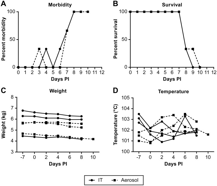
Nipah virus (NiV) is a paramyxovirus (genus Henipavirus) that emerged in the late 1990s in Malaysia and has since been identified as the cause of sporadic outbreaks of severe febrile disease in Bangladesh and India. NiV infection is frequently associated with severe respiratory or neurological disease in infected humans with transmission to humans through inhalation, contact or consumption of NiV contaminated foods. In the work presented here, the development of disease was investigated in the African Green Monkey (AGM) model following intratracheal (IT) and, for the first time, small-particle aerosol administration of NiV. This study utilized computed tomography (CT) and magnetic resonance imaging (MRI) to temporally assess disease progression. The host immune response and changes in immune cell populations over the course of disease were also evaluated. This study found that IT and small-particle administration of NiV caused similar disease progression, but that IT inoculation induced significant congestion in the lungs while disease following small-particle aerosol inoculation was largely confined to the lower respiratory tract. Quantitative assessment of changes in lung volume found up to a 45% loss in IT inoculated animals. None of the subjects in this study developed overt neurological disease, a finding that was supported by MRI analysis. The development of neutralizing antibodies was not apparent over the 8-10 day course of disease, but changes in cytokine response in all animals and activated CD8+ T cell numbers suggest the onset of cell-mediated immunity. These studies demonstrate that IT and small-particle aerosol infection with NiV in the AGM model leads to a severe respiratory disease devoid of neurological indications. This work also suggests that extending the disease course or minimizing the impact of the respiratory component is critical to developing a model that has a neurological component and more accurately reflects the human condition.
尼帕病毒(NiV)是一种副粘病毒(亨尼帕病毒属),于20世纪90年代末在马来西亚出现,此后被确定为孟加拉国和印度零星爆发严重发热疾病的病因。尼帕病毒感染在受感染人类中常与严重的呼吸道或神经系统疾病相关,可通过吸入、接触或食用受尼帕病毒污染的食物传播给人类。在本文介绍的研究中,在非洲绿猴(AGM)模型中,通过气管内(IT)以及首次通过小颗粒气溶胶方式接种尼帕病毒,研究了疾病的发展情况。本研究利用计算机断层扫描(CT)和磁共振成像(MRI)对疾病进展进行了时间上的评估。还评估了疾病过程中宿主的免疫反应以及免疫细胞群体的变化。本研究发现,通过IT和小颗粒方式接种尼帕病毒导致了相似的疾病进展,但IT接种在肺部引起了明显的充血,而小颗粒气溶胶接种后的疾病主要局限于下呼吸道。对肺容积变化的定量评估发现,IT接种的动物肺容积损失高达45%。本研究中没有受试者出现明显的神经系统疾病,这一发现得到了MRI分析的支持。在疾病的8 - 10天病程中,中和抗体的产生并不明显,但所有动物细胞因子反应的变化以及活化的CD8 + T细胞数量表明细胞介导免疫开始。这些研究表明,在AGM模型中通过IT和小颗粒气溶胶感染尼帕病毒会导致严重的呼吸道疾病,且无神经系统症状。这项工作还表明,延长病程或尽量减少呼吸道成分的影响对于建立一个具有神经系统成分并能更准确反映人类病情的模型至关重要。