• 文献检索
  • 文档翻译
  • 深度研究
  • 学术资讯
  • Suppr Zotero 插件Zotero 插件
  • 邀请有礼
  • 套餐&价格
  • 历史记录
应用&插件
Suppr Zotero 插件Zotero 插件浏览器插件Mac 客户端Windows 客户端微信小程序
定价
高级版会员购买积分包购买API积分包
服务
文献检索文档翻译深度研究API 文档MCP 服务
关于我们
关于 Suppr公司介绍联系我们用户协议隐私条款
关注我们

Suppr 超能文献

核心技术专利:CN118964589B侵权必究
粤ICP备2023148730 号-1Suppr @ 2026

文献检索

告别复杂PubMed语法,用中文像聊天一样搜索,搜遍4000万医学文献。AI智能推荐,让科研检索更轻松。

立即免费搜索

文件翻译

保留排版,准确专业,支持PDF/Word/PPT等文件格式,支持 12+语言互译。

免费翻译文档

深度研究

AI帮你快速写综述,25分钟生成高质量综述,智能提取关键信息,辅助科研写作。

立即免费体验

相干 X 射线衍射成像技术对哺乳动物线粒体的可视化研究。

Visualization of a Mammalian Mitochondrion by Coherent X-ray Diffractive Imaging.

机构信息

Department of Physics and Photon Science & School of Materials Science and Engineering, Gwangju Institute of Science and Technology, Gwangju, 61005, Korea.

European XFEL, Schenefeld, 22869, Germany.

出版信息

Sci Rep. 2017 May 12;7(1):1850. doi: 10.1038/s41598-017-01833-x.

DOI:10.1038/s41598-017-01833-x
PMID:28500280
原文链接:https://pmc.ncbi.nlm.nih.gov/articles/PMC5431869/
Abstract

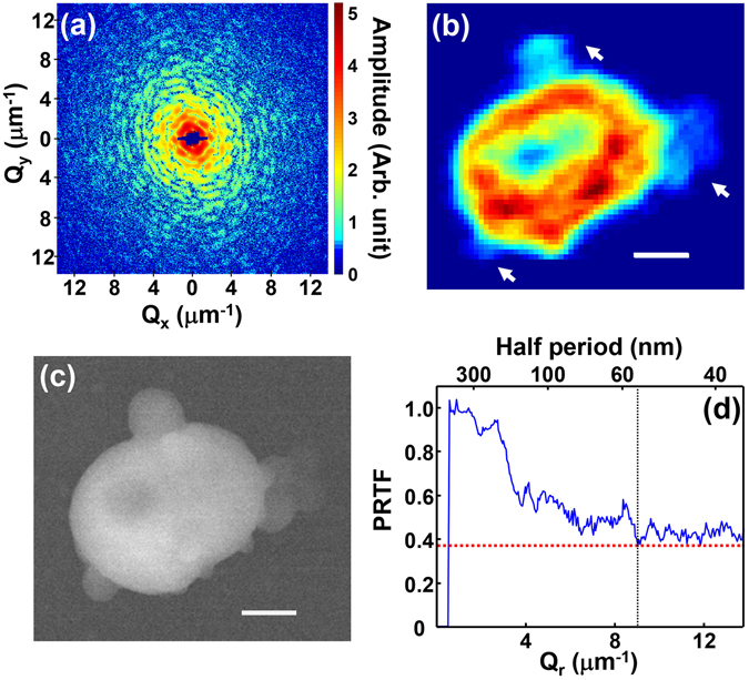
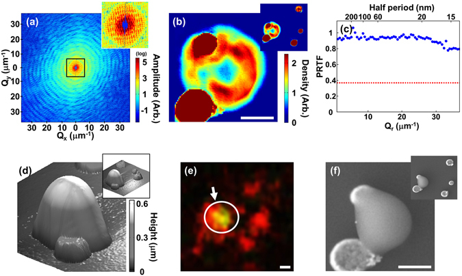
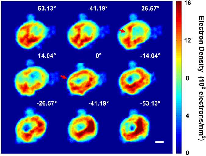
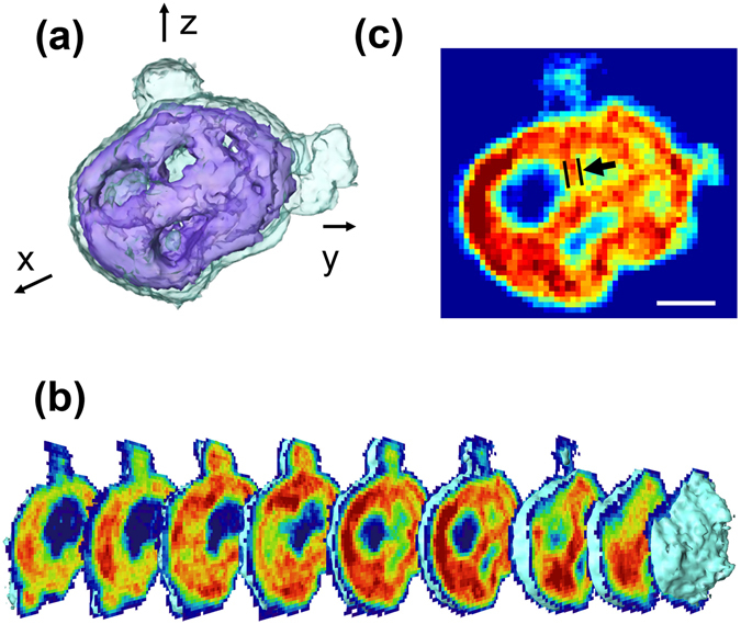
We report a three dimensional (3D) quantitative visualization of a mammalian mitochondrion by coherent x-ray diffractive imaging (CXDI) using synchrotron radiation. The internal structures of a mitochondrion from a mouse embryonic fibroblast cell line (NIH3T3) were visualized by tomographic imaging at approximately 60 nm resolution without the need for sectioning or staining. The overall structure consisted of a high electron density region, composed of the outer and inner membranes and the cristae cluster, which enclosed the lower density mitochondrial matrix. The average mass density of the mitochondrion was about 1.36 g/cm. Sectioned images of the cristae reveal that they have neither a baffle nor septa shape but were instead irregular. In addition, a high resolution, about 14 nm, 2D projection image was captured of a similar mitochondrion with the aid of strongly scattering Au reference objects. Obtaining 3D images at this improved resolution will allow CXDI to be an effective and nondestructive method for investigating the innate structure of mitochondria and other important life supporting organelles.

摘要

我们通过使用同步辐射的相干 X 射线衍射成像(CXDI)报告了哺乳动物线粒体的三维(3D)定量可视化。通过层析成像,在无需切片或染色的情况下,以约 60nm 的分辨率对来自小鼠胚胎成纤维细胞系(NIH3T3)的线粒体的内部结构进行了可视化。整体结构由一个高电子密度区域组成,该区域由外膜、内膜和嵴簇组成,这些结构包围着密度较低的线粒体基质。线粒体的平均质量密度约为 1.36g/cm³。对嵴的切片图像表明,它们既没有隔板也没有隔片形状,而是不规则的。此外,借助强烈散射的 Au 参考物体,还捕获了类似线粒体的高分辨率(约 14nm)2D 投影图像。以这种提高的分辨率获得 3D 图像将使 CXDI 成为一种有效且无损的方法,用于研究线粒体和其他重要生命支持细胞器的固有结构。

https://cdn.ncbi.nlm.nih.gov/pmc/blobs/08f9/5431869/1426097cb5e4/41598_2017_1833_Fig4_HTML.jpg
https://cdn.ncbi.nlm.nih.gov/pmc/blobs/08f9/5431869/81be7692517b/41598_2017_1833_Fig1_HTML.jpg
https://cdn.ncbi.nlm.nih.gov/pmc/blobs/08f9/5431869/dbfda586779a/41598_2017_1833_Fig2_HTML.jpg
https://cdn.ncbi.nlm.nih.gov/pmc/blobs/08f9/5431869/51f9045619db/41598_2017_1833_Fig3_HTML.jpg
https://cdn.ncbi.nlm.nih.gov/pmc/blobs/08f9/5431869/1426097cb5e4/41598_2017_1833_Fig4_HTML.jpg

相似文献

1
Visualization of a Mammalian Mitochondrion by Coherent X-ray Diffractive Imaging.相干 X 射线衍射成像技术对哺乳动物线粒体的可视化研究。
Sci Rep. 2017 May 12;7(1):1850. doi: 10.1038/s41598-017-01833-x.
2
Coherent X-Ray Diffraction Imaging of Chloroplasts from Cyanidioschyzon merolae by Using X-Ray Free Electron Laser.利用X射线自由电子激光对嗜热栖热放线菌叶绿体进行相干X射线衍射成像
Plant Cell Physiol. 2015 Jul;56(7):1272-86. doi: 10.1093/pcp/pcv032. Epub 2015 Mar 5.
3
Coherent diffractive imaging of biological samples at synchrotron and free electron laser facilities.同步辐射和自由电子激光设施下生物样本的相干衍射成像。
J Biotechnol. 2010 Sep 15;149(4):229-37. doi: 10.1016/j.jbiotec.2010.01.024. Epub 2010 Feb 10.
4
Resolution enhancement in coherent x-ray diffraction imaging by overcoming instrumental noise.通过克服仪器噪声提高相干X射线衍射成像的分辨率
Opt Express. 2014 Nov 17;22(23):29161-9. doi: 10.1364/OE.22.029161.
5
Internal structure of an intact Convallaria majalis pollen grain observed with X-ray Fresnel coherent diffractive imaging.用X射线菲涅耳相干衍射成像观察到的完整铃兰花粉粒的内部结构。
Opt Express. 2012 Nov 19;20(24):26778-85. doi: 10.1364/OE.20.026778.
6
Tomography of a Cryo-immobilized Yeast Cell Using Ptychographic Coherent X-Ray Diffractive Imaging.使用叠层相干X射线衍射成像技术对冷冻固定酵母细胞进行断层扫描。
Biophys J. 2015 Nov 3;109(9):1986-95. doi: 10.1016/j.bpj.2015.08.047.
7
Electron tomography of neuronal mitochondria: three-dimensional structure and organization of cristae and membrane contacts.神经元线粒体的电子断层扫描:嵴和膜接触的三维结构与组织
J Struct Biol. 1997 Aug;119(3):260-72. doi: 10.1006/jsbi.1997.3885.
8
X-Ray Structural Analysis of Single Adult Cardiomyocytes: Tomographic Imaging and Microdiffraction.单个成年心肌细胞的X射线结构分析:断层成像与微衍射
Biophys J. 2020 Oct 6;119(7):1309-1323. doi: 10.1016/j.bpj.2020.08.019. Epub 2020 Aug 28.
9
Three-dimensional imaging of mitochondrial cristae complexity using cryo-soft X-ray tomography.利用冷冻软 X 射线断层成像技术对线粒体嵴复杂度进行三维成像。
Sci Rep. 2020 Dec 3;10(1):21045. doi: 10.1038/s41598-020-78150-3.
10
Coherent X-ray diffractive imaging of protein crystals.蛋白质晶体的相干X射线衍射成像
J Synchrotron Radiat. 2008 Nov;15(Pt 6):576-83. doi: 10.1107/S0909049508029439. Epub 2008 Oct 3.

引用本文的文献

1
Simultaneous targeting and suppression of heat shock protein 60 to overcome heat resistance and induce mitochondrial death of tumor cells in photothermal immunotherapy.在光热免疫疗法中同时靶向并抑制热休克蛋白60以克服热抗性并诱导肿瘤细胞的线粒体死亡。
Mater Today Bio. 2024 Sep 29;29:101282. doi: 10.1016/j.mtbio.2024.101282. eCollection 2024 Dec.
2
Expected resolution limits of x-ray free-electron laser single-particle imaging for realistic source and detector properties.基于实际光源和探测器特性的X射线自由电子激光单粒子成像的预期分辨率极限。
Struct Dyn. 2022 Nov 16;9(6):064101. doi: 10.1063/4.0000169. eCollection 2022 Nov.
3

本文引用的文献

1
Enhancing resolution in coherent x-ray diffraction imaging.提高相干X射线衍射成像的分辨率。
J Phys Condens Matter. 2016 Dec 14;28(49):493001. doi: 10.1088/0953-8984/28/49/493001. Epub 2016 Oct 18.
2
Three-dimensional coherent X-ray diffractive imaging of whole frozen-hydrated cells.三维相干 X 射线衍射成像技术对整个冷冻水合细胞的研究
IUCrJ. 2015 Aug 20;2(Pt 5):575-83. doi: 10.1107/S205225251501235X. eCollection 2015 Sep 1.
3
Resolution enhancement in coherent x-ray diffraction imaging by overcoming instrumental noise.
Advanced Light Source Analytical Techniques for Exploring the Biological Behavior and Fate of Nanomedicines.
用于探索纳米药物生物学行为和归宿的先进光源分析技术
ACS Cent Sci. 2022 Aug 24;8(8):1063-1080. doi: 10.1021/acscentsci.2c00680. Epub 2022 Aug 1.
4
Electromagnetic Dosimetry for Isolated Mitochondria Exposed to Near-Infrared Continuous-Wave Illumination in Photobiomodulation Experiments.在光生物调节实验中,对近红外连续波辐照下的分离线粒体进行电磁剂量测定。
Bioelectromagnetics. 2021 Jul;42(5):384-397. doi: 10.1002/bem.22342. Epub 2021 May 18.
5
Three-dimensional imaging of mitochondrial cristae complexity using cryo-soft X-ray tomography.利用冷冻软 X 射线断层成像技术对线粒体嵴复杂度进行三维成像。
Sci Rep. 2020 Dec 3;10(1):21045. doi: 10.1038/s41598-020-78150-3.
6
Laser-induced metastable mixed phase of AuNi nanoparticles: a coherent X-ray diffraction imaging study.激光诱导的金镍纳米颗粒亚稳混合相:相干X射线衍射成像研究
J Synchrotron Radiat. 2020 May 1;27(Pt 3):725-729. doi: 10.1107/S1600577520001617. Epub 2020 Mar 31.
7
Coherent diffractive imaging of microtubules using an X-ray laser.使用 X 射线激光对微管进行相干衍射成像。
Nat Commun. 2019 Jun 13;10(1):2589. doi: 10.1038/s41467-019-10448-x.
8
Spectral comparisons of mammalian cells and intact organelles by solid-state NMR.固态 NMR 技术对哺乳动物细胞和完整细胞器的光谱比较。
J Struct Biol. 2019 Apr 1;206(1):49-54. doi: 10.1016/j.jsb.2018.05.007. Epub 2018 May 30.
通过克服仪器噪声提高相干X射线衍射成像的分辨率
Opt Express. 2014 Nov 17;22(23):29161-9. doi: 10.1364/OE.22.029161.
4
Analytic 3D imaging of mammalian nucleus at nanoscale using coherent x-rays and optical fluorescence microscopy.利用相干X射线和光学荧光显微镜对哺乳动物细胞核进行纳米级分析3D成像。
Biophys J. 2014 Sep 2;107(5):1074-1081. doi: 10.1016/j.bpj.2014.07.028.
5
Macromolecular structures probed by combining single-shot free-electron laser diffraction with synchrotron coherent X-ray imaging.利用单次自由电子激光衍射与同步辐射相干 X 射线成像技术探测大分子结构。
Nat Commun. 2014 May 2;5:3798. doi: 10.1038/ncomms4798.
6
Self-terminating diffraction gates femtosecond X-ray nanocrystallography measurements.自终止衍射门飞秒X射线纳米晶体学测量。
Nat Photonics. 2012;6:35-40. doi: 10.1038/nphoton.2011.297.
7
Mitochondrial network morphology: building an integrative, geometrical view.线粒体网络形态:构建一个综合的、几何的视角。
BMC Biol. 2013 Jun 24;11:71. doi: 10.1186/1741-7007-11-71.
8
Imaging fully hydrated whole cells by coherent x-ray diffraction microscopy.利用相干 X 射线衍射显微镜对完全水合的整个细胞进行成像。
Phys Rev Lett. 2013 Mar 1;110(9):098103. doi: 10.1103/PhysRevLett.110.098103. Epub 2013 Feb 28.
9
Intracellular label-free gold nanorods imaging with photoacoustic microscopy.利用光声显微镜对细胞内无标记金纳米棒进行成像。
Opt Express. 2012 Apr 23;20(9):10370-5. doi: 10.1364/OE.20.010370.
10
Electron tomography at 2.4-ångström resolution.2.4 埃电子断层成像术。
Nature. 2012 Mar 21;483(7390):444-7. doi: 10.1038/nature10934.