Isomura Shuichi, Hashimoto Ryota, Nakamura Motoaki, Hirano Yoji, Yamashita Fumio, Jimbo Shin, Yamamori Hidenaga, Fujimoto Michiko, Yasuda Yuka, Mears Ryan P, Onitsuka Toshiaki
Department of Neuropsychiatry, Graduate School of Medical Sciences, Kyushu University, 3-1-1, Maidashi, Higashiku, Fukuoka, Japan.
Molecular Research Center for Children's Mental Development, United Graduate School of Child Development, Osaka University, Osaka, Japan.
NPJ Schizophr. 2017 Jan 12;3:3. doi: 10.1038/s41537-016-0008-y. eCollection 2017.
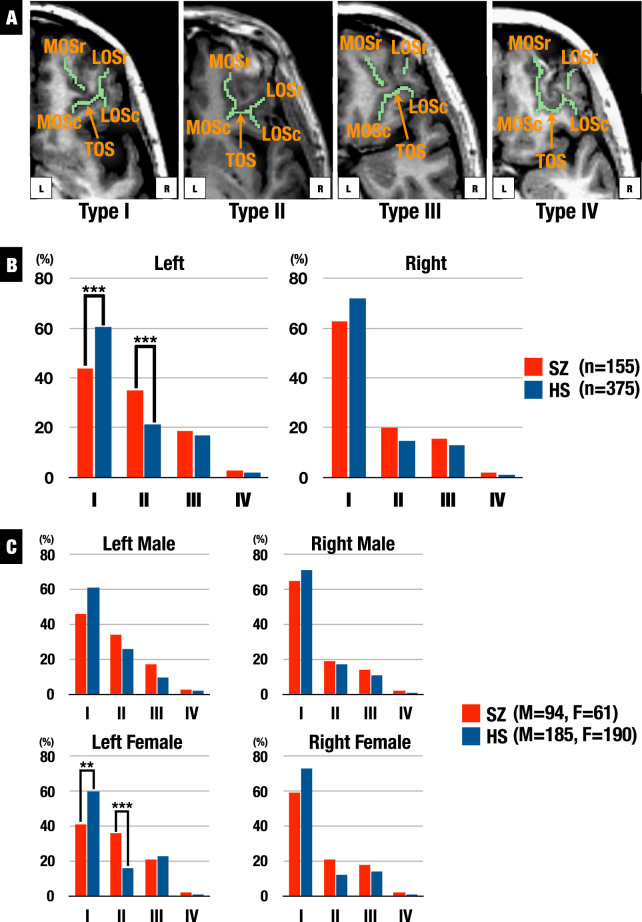
Abnormalities in prenatal brain development contribute to schizophrenia vulnerability. Orbitofrontal cortex sulcogyral patterns are largely determined during prenatal development, and four types of orbitofrontal cortex sulcogyral patterns have been classified in humans. Altered orbitofrontal cortex patterns have been reported in individuals with schizophrenia using magnetic resonance imaging; however, sample sizes of previous studies were small-medium effects for detection, and gender manifestation for orbitofrontal cortex sulcogyral patterns is unclear. The present study investigated orbitofrontal cortex patterns of 155 patients with schizophrenia and 375 healthy subjects. The orbitofrontal cortex sulcogyral pattern distributions of schizophrenia were significantly different compared with healthy subjects in the left hemisphere ( = 14.55, = 0.002). In female schizophrenia, post-hoc analyses revealed significantly decreased Type I expression ( = 6.76, = 0.009) and increased Type II expression ( = 11.56, = 0.001) in the left hemisphere. The present study suggested that female schizophrenia showed altered orbitofrontal cortex patterns in the left hemisphere, which may be related to neurodevelopmental abnormality.
产前大脑发育异常会导致精神分裂症易感性。眶额皮质沟回模式在很大程度上是在产前发育期间确定的,并且在人类中已分类出四种眶额皮质沟回模式。使用磁共振成像已报道精神分裂症患者存在眶额皮质模式改变;然而,先前研究的样本量对检测来说是中效的,并且眶额皮质沟回模式的性别表现尚不清楚。本研究调查了155例精神分裂症患者和375名健康受试者的眶额皮质模式。精神分裂症患者的眶额皮质沟回模式分布与健康受试者相比,在左半球有显著差异(=14.55,=0.002)。在女性精神分裂症患者中,事后分析显示左半球I型表达显著降低(=6.76,=0.009),II型表达增加(=11.56,=0.001)。本研究表明,女性精神分裂症患者左半球的眶额皮质模式发生改变,这可能与神经发育异常有关。