Department of Life Science, National Taiwan Normal University, Taipei, 10677, Taiwan.
Department of Medical Imaging, Taipei TzuChi Hospital, The Buddhist TzuChi Medical Foundation, New Taipei City, 23142, Taiwan.
Sci Rep. 2017 Jun 15;7(1):3587. doi: 10.1038/s41598-017-03863-x.
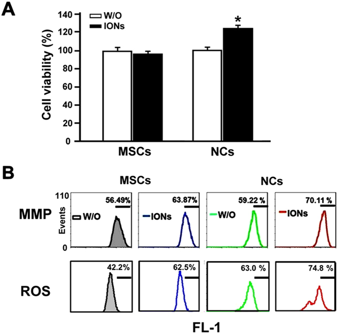
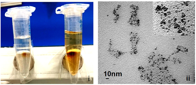
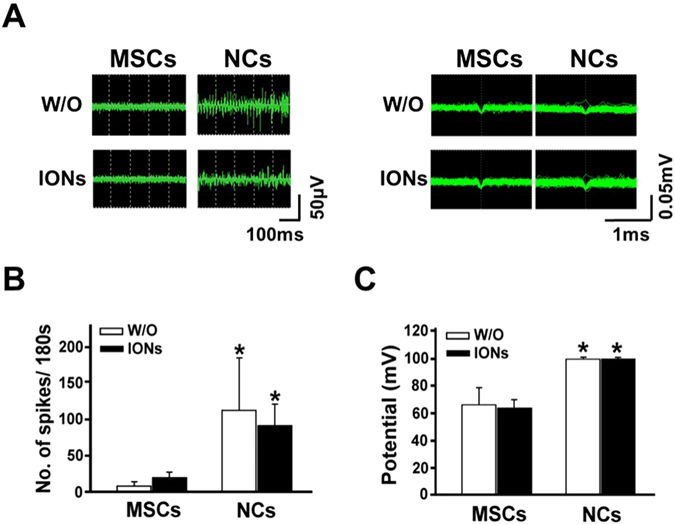
The aim of the current study was to develop an iron oxide nanoparticle (ION) labelling and magnetic resonance imaging (MRI)-based protocol to allow visualization of the differentiation process of mesenchymal stem cells (MSCs) into neural-like cells (NCs) in vitro. Ferucarbotran, a clinically available ION, which can be visualized under MRI, is used for tracking cells implanted in vivo. The NCs were verified morphologically and histologically by light microscopy, and their functions were verified by measuring their action potentials. Conformational conversion of axon-like structures was observed under light microscopy. These NCs exhibited frequent, active action potentials compared with cells that did not undergo neural differentiation. The labelling of ION had no influence on the morphological and functional differentiation capacity of the MSCs. We conclude that the MSCs that were differentiated into NCs exhibited in vitro activity potential firing and may be used to replace damaged neurons.
本研究旨在开发一种基于氧化铁纳米颗粒(ION)标记和磁共振成像(MRI)的方案,以可视化间充质干细胞(MSCs)在体外向神经样细胞(NCs)分化的过程。Ferucarbotran 是一种临床可用的 ION,可以在 MRI 下可视化,用于跟踪体内植入的细胞。通过光学显微镜验证 NCs 的形态和组织学,并通过测量动作电位来验证其功能。在光学显微镜下观察到类似轴突的结构的构象转换。与未经历神经分化的细胞相比,这些 NCs 表现出频繁、活跃的动作电位。ION 的标记对 MSC 的形态和功能分化能力没有影响。我们得出结论,分化为 NC 的 MSC 表现出体外活动电位发射,可用于替代受损神经元。