Suinesiaputra Avan, Sanghvi Mihir M, Aung Nay, Paiva Jose Miguel, Zemrak Filip, Fung Kenneth, Lukaschuk Elena, Lee Aaron M, Carapella Valentina, Kim Young Jin, Francis Jane, Piechnik Stefan K, Neubauer Stefan, Greiser Andreas, Jolly Marie-Pierre, Hayes Carmel, Young Alistair A, Petersen Steffen E
Department of Anatomy and Medical Imaging, Faculty of Medical and Health Sciences, University of Auckland, 85 Park Road, Auckland, 1142, New Zealand.
William Harvey Research Institute, NIHR Cardiovascular Biomedical Research Centre at Barts, Queen Mary University of London, Charterhouse Square, London, EC1M 6BQ, UK.
Int J Cardiovasc Imaging. 2018 Feb;34(2):281-291. doi: 10.1007/s10554-017-1225-9. Epub 2017 Aug 23.
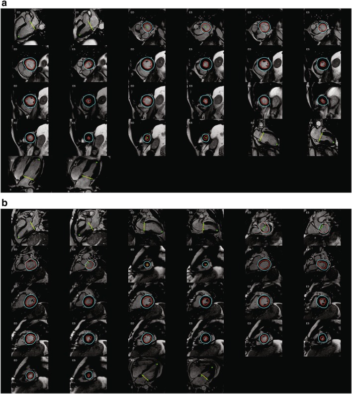
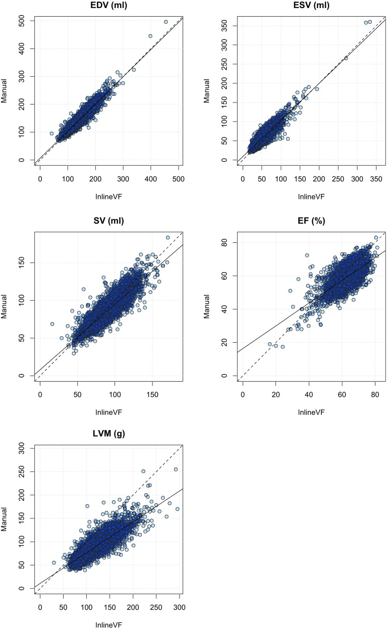
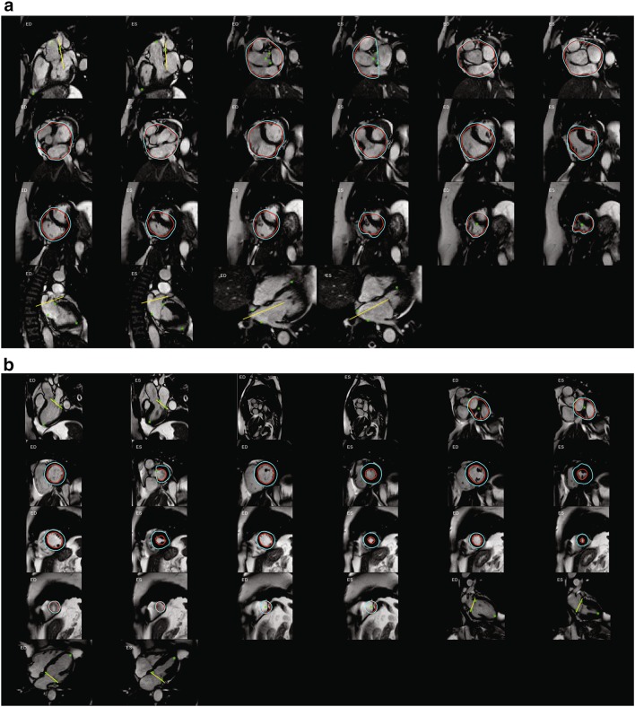
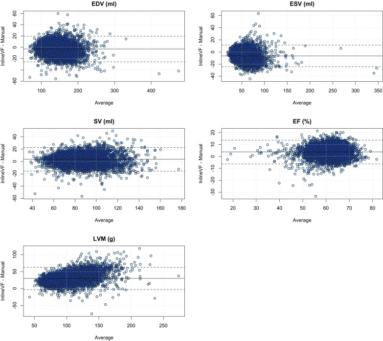
UK Biobank, a large cohort study, plans to acquire 100,000 cardiac MRI studies by 2020. Although fully-automated left ventricular (LV) analysis was performed in the original acquisition, this was not designed for unsupervised incorporation into epidemiological studies. We sought to evaluate automated LV mass and volume (Siemens syngo InlineVF versions D13A and E11C), against manual analysis in a substantial sub-cohort of UK Biobank participants. Eight readers from two centers, trained to give consistent results, manually analyzed 4874 UK Biobank cases for LV end-diastolic volume (EDV), end-systolic volume (ESV), stroke volume (SV), ejection fraction (EF) and LV mass (LVM). Agreement between manual and InlineVF automated analyses were evaluated using Bland-Altman analysis and the intra-class correlation coefficient (ICC). Tenfold cross-validation was used to establish a linear regression calibration between manual and InlineVF results. InlineVF D13A returned results in 4423 cases, whereas InlineVF E11C returned results in 4775 cases and also reported LVM. Rapid visual assessment of the E11C results found 178 cases (3.7%) with grossly misplaced contours or landmarks. In the remaining 4597 cases, LV function showed good agreement: ESV -6.4 ± 9.0 ml, 0.853 (mean ± SD of the differences, ICC) EDV -3.0 ± 11.6 ml, 0.937; SV 3.4 ± 9.8 ml, 0.855; and EF 3.5 ± 5.1%, 0.586. Although LV mass was consistently overestimated (29.9 ± 17.0 g, 0.534) due to larger epicardial contours on all slices, linear regression could be used to correct the bias and improve accuracy. Automated InlineVF results can be used for case-control studies in UK Biobank, provided visual quality control and linear bias correction are performed. Improvements between InlineVF D13A and InlineVF E11C show the field is rapidly advancing, with further improvements expected in the near future.
英国生物银行是一项大型队列研究,计划到2020年获取10万份心脏磁共振成像研究资料。尽管在最初采集时进行了全自动左心室(LV)分析,但这并非为无监督地纳入流行病学研究而设计。我们试图在英国生物银行参与者的一个相当大的子队列中,将自动LV质量和容积(西门子syngo InlineVF版本D13A和E11C)与手动分析进行对比评估。来自两个中心的8名读者经过培训以给出一致的结果,他们手动分析了4874例英国生物银行病例的左心室舒张末期容积(EDV)、收缩末期容积(ESV)、每搏输出量(SV)、射血分数(EF)和左心室质量(LVM)。使用Bland-Altman分析和组内相关系数(ICC)评估手动分析和InlineVF自动分析之间的一致性。采用十折交叉验证来建立手动分析和InlineVF结果之间的线性回归校准。InlineVF D13A在4423例病例中返回了结果,而InlineVF E11C在4775例病例中返回了结果并且还报告了LVM。对E11C结果的快速视觉评估发现178例(3.7%)病例的轮廓或地标严重错位。在其余4597例病例中,左心室功能显示出良好的一致性:ESV -6.4±9.0毫升,差异的均值±标准差为0.853(ICC);EDV -3.0±11.6毫升,0.937;SV 3.4±9.8毫升,0.855;EF 3.5±5.1%,0.586。尽管由于所有切片上的心外膜轮廓较大,LV质量一直被高估(29.9±17.0克,0.534),但可以使用线性回归来校正偏差并提高准确性。只要进行视觉质量控制和线性偏差校正,自动InlineVF结果可用于英国生物银行的病例对照研究。InlineVF D13A和InlineVF E11C之间的改进表明该领域正在迅速发展,预计在不久的将来会有进一步的改进。