Iqbal Sakib, Rashid Mohammad H, Arbab Ali S, Khan Mujibur
Mechanical Engineering, Georgia Southern University, Statesboro, GA.
Georgia Cancer Center; Augusta University, Augusta, GA.
J Biomed Nanotechnol. 2017 Apr;13(4):355-366. doi: 10.1166/jbn.2017.2353.
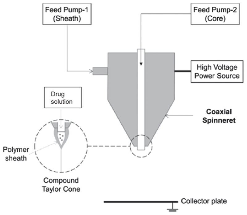
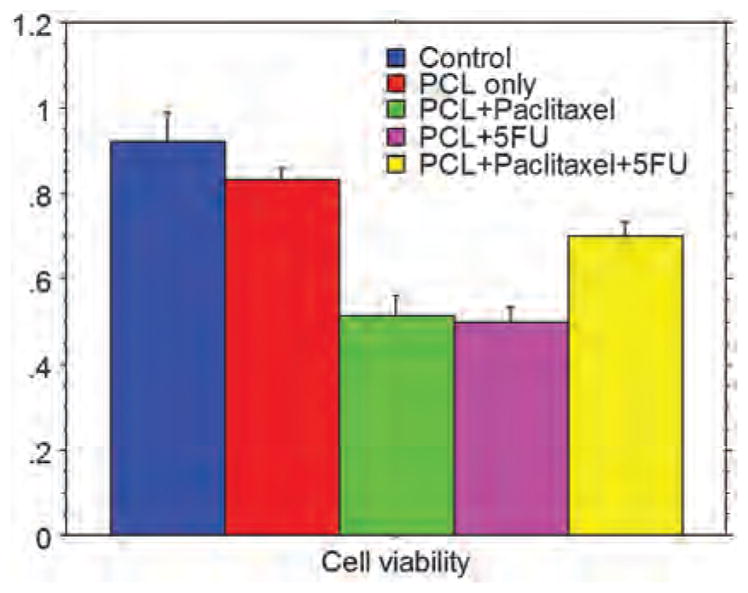
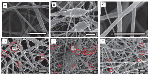
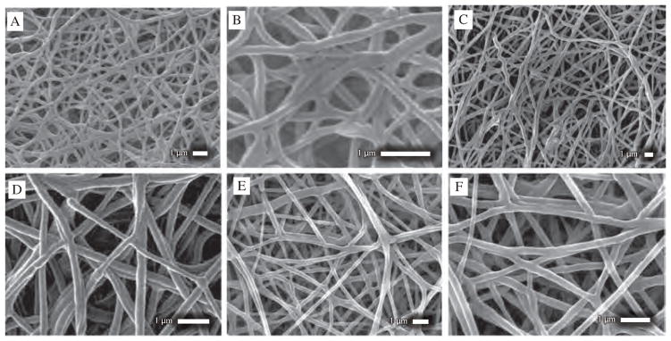
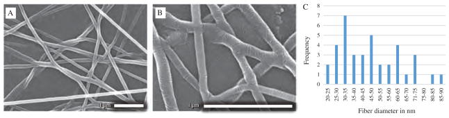
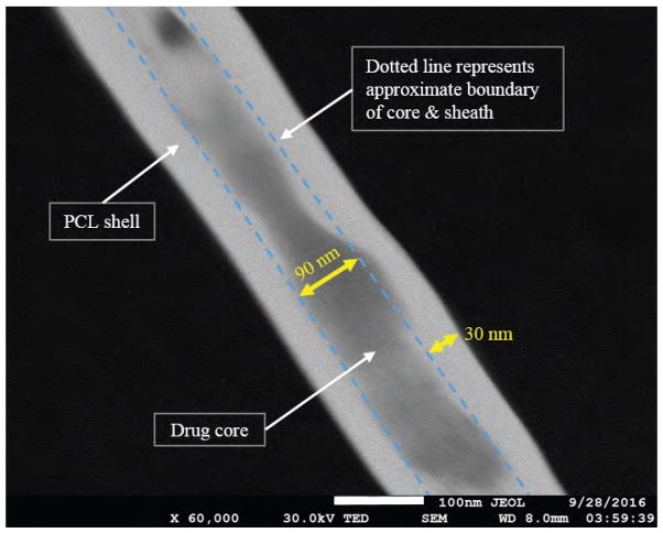
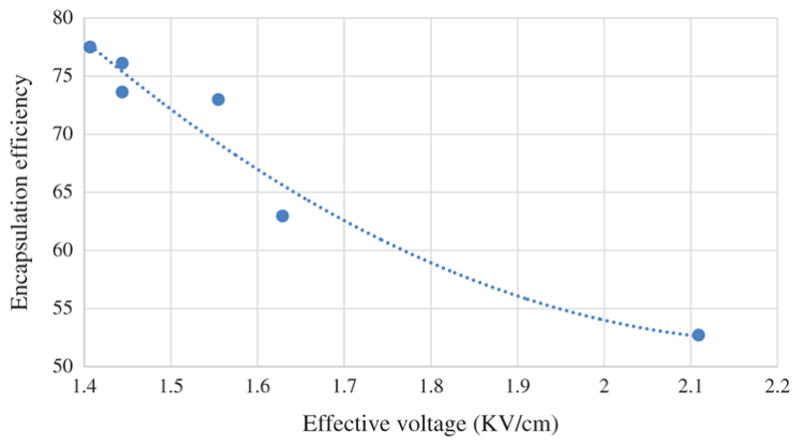
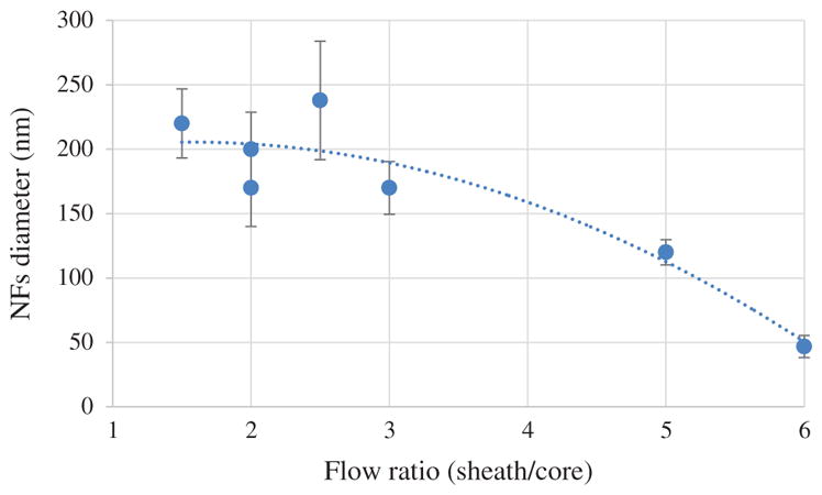
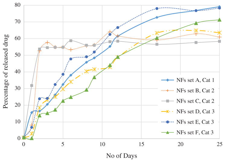
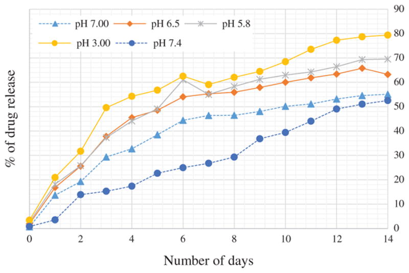
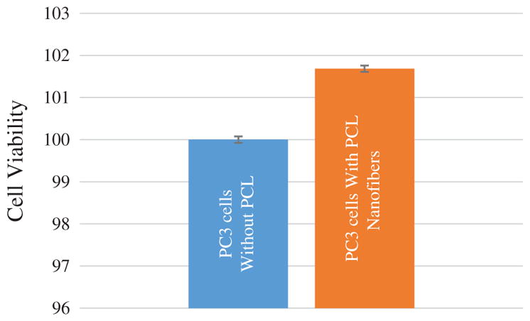
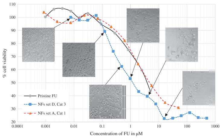
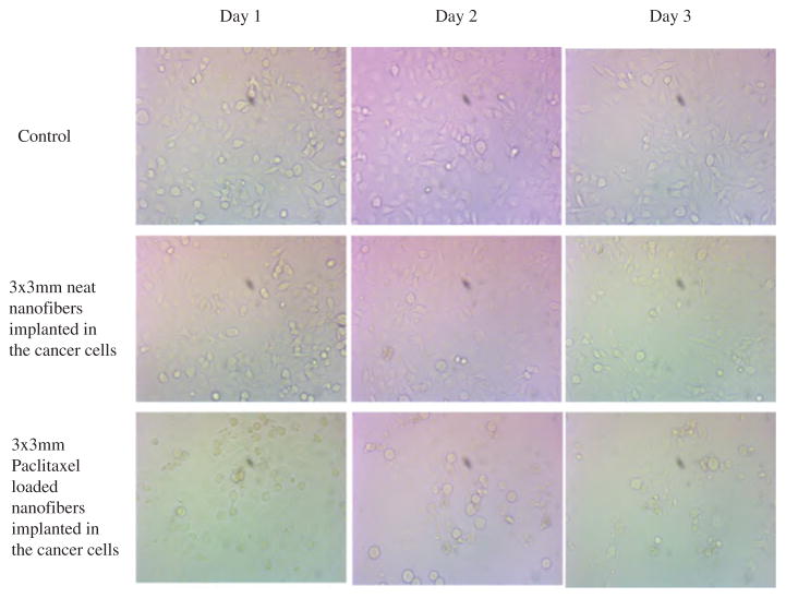
We report a continuous nanoscale encapsulation of cancer drugs 5-Fluorouracil (FU) and Paclitaxel into biocompatible polycaprolactone (PCL) nanofibers (NFs) using core-sheath electrospinning process. A high potential electric field of 19-23.2 kV was used to draw a compound solution jet from a specialized coaxial spinneret. Using of DMF in both core and Sheath resulted in NFs within 50-160 nm along with large beaded structures. Addition of Trichloromethane (TCM) or Trifluoroethanol (TFE) in sheath turned NFs in more uniform and thin fiber structure. The diameter range for paclitaxel encapsulated fibers was 22-90 nm with encapsulation efficiency of 77.5% and the amount of drug was only 4 to 5% of sheath polymer. Addition of PVA within core resulted drug nanocrystal formation outside of sheath and poor encapsulation efficiency (52%) with rapid initial release (52-53%) in first 3 days. Drug release test of NFs in different pH exhibited increase of release rate with the decrease of media pH. In-vitro cell viability test with FU encapsulated NFs in human prostatic cancer PC3 cells exhibited 38% alive cells at 5 μM concentration while in pristine FU 43% cells were alive. Paclitaxel encapsulated NFs with breast cancer cells also exhibited increased efficacy in comparison to pristine anticancer drugs. Continuous decrease of cell density indicated the slow release of cancer drugs from the NFs. Both PCL+Paclitaxel and PCL+5FU treated conditions caused breast cancer cell death between 40% to 50%.
我们报道了使用核壳电纺工艺将癌症药物5-氟尿嘧啶(FU)和紫杉醇连续纳米级封装到生物相容性聚己内酯(PCL)纳米纤维(NFs)中。使用19 - 23.2 kV的高电位电场从专门的同轴喷丝头中拉出复合溶液射流。在核层和壳层中都使用二甲基甲酰胺(DMF),得到了50 - 160 nm范围内的纳米纤维以及大的珠状结构。在壳层中添加三氯甲烷(TCM)或三氟乙醇(TFE),使纳米纤维具有更均匀和更细的纤维结构。封装紫杉醇的纤维直径范围为22 - 90 nm,封装效率为77.5%,药物含量仅为壳层聚合物的4%至5%。在核层中添加聚乙烯醇(PVA)导致在壳层外形成药物纳米晶体,封装效率低(52%),在前3天有快速的初始释放(52 - 53%)。不同pH值下纳米纤维的药物释放测试表明,随着培养基pH值的降低,释放速率增加。用封装了FU的纳米纤维对人前列腺癌PC3细胞进行体外细胞活力测试,在5 μM浓度下有38%的活细胞,而在原始FU中43%的细胞存活。与原始抗癌药物相比,封装紫杉醇的纳米纤维对乳腺癌细胞也表现出更高的疗效。细胞密度的持续降低表明癌症药物从纳米纤维中缓慢释放。PCL + 紫杉醇和PCL + 5FU处理条件均导致40%至50%的乳腺癌细胞死亡。