Pixley Sarah K, Hopkins Tracy M, Little Kevin J, Hom David B
Department of Molecular and Cellular Physiology (S.K.P., T.M.H.) Cincinnati Children's Hospital Medical Center Cincinnati Ohio U.S.A.
Pediatric Hand and Upper Extremity Center (K.J.L.), Cincinnati Children's Hospital Medical Center Cincinnati Ohio U.S.A.
Laryngoscope Investig Otolaryngol. 2016 Nov 14;1(6):185-190. doi: 10.1002/lio2.41. eCollection 2016 Dec.
Hollow nerve conduits made of natural or synthetic biomaterials are used clinically to aid regeneration of peripheral nerves damaged by trauma or disease. To support healing, conduit lumen patency must be maintained until recovery occurs. New methods to study conduit structural integrity would provide an important means to optimize conduits in preclinical studies. We explored a novel combined technique to examine structural integrity of two types of nerve conduits after in vivo healing.
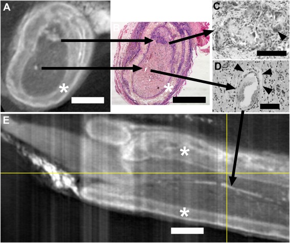
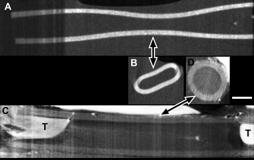
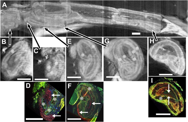
Micro-CT imaging with iodine contrast was combined with histological analysis to examine two different nerve conduits after in vivo nerve reconstruction in rats.
Sciatic nerve gaps in adult Lewis rats were reconstructed with poly(caprolactone) (PCL, 1.6 cm gap, 14-week survival) or silicone (1 cm gap, 6-week survival) conduits (N = 12 total). Conduits with regenerating tissues were imaged by micro-CT with iodine contrast and compared to the histology (hematoxylin and eosin, immunostaining for axons) of regenerated tissues after iodine removal.
PCL nerve conduits showed extensive breakage throughout their length, but all showed successful nerve growth through the conduits. The silicone conduits remained intact, although significant constriction was uniquely detected by micro-CT, with 1 of 6 animals showing incomplete tissue regeneration.
Micro-CT with iodine contrast offers a unique and valuable means to determine 3D structural integrity of nerve conduits and nerve healing following reconstruction. Furthermore, this paper shows that even if conduit compression and degradation occur, nerve regeneration can still take place.
由天然或合成生物材料制成的中空神经导管在临床上用于辅助因创伤或疾病受损的周围神经再生。为了促进愈合,在恢复之前必须保持导管腔的通畅。研究导管结构完整性的新方法将为临床前研究中优化导管提供重要手段。我们探索了一种新颖的联合技术,以检查两种类型的神经导管在体内愈合后的结构完整性。
将含碘造影剂的微型计算机断层扫描(Micro-CT)成像与组织学分析相结合,以检查大鼠体内神经重建后两种不同的神经导管。
用聚己内酯(PCL,间隙1.6厘米,存活14周)或硅胶(间隙1厘米,存活6周)导管重建成年Lewis大鼠的坐骨神经间隙(总共N = 12)。对含有再生组织的导管进行含碘造影剂的微型计算机断层扫描成像,并与去除碘后再生组织的组织学(苏木精和伊红染色、轴突免疫染色)进行比较。
PCL神经导管在其整个长度上显示出广泛的破损,但所有导管都显示出神经成功地通过导管生长。硅胶导管保持完整,尽管微型计算机断层扫描独特地检测到明显的狭窄,6只动物中有1只显示组织再生不完全。
含碘造影剂的微型计算机断层扫描提供了一种独特且有价值的方法,用于确定神经导管的三维结构完整性以及重建后的神经愈合情况。此外,本文表明即使发生导管压缩和降解,神经再生仍可发生。