Division of Clinical Geriatrics, Department of Neurobiology, Care Sciences and Society, Karolinska Institute, Stockholm, Sweden.
Clinical Memory Research Unit, Department of Clinical Sciences Malmö, Lund University, Lund, Sweden.
Cereb Cortex. 2018 Jan 1;28(1):340-349. doi: 10.1093/cercor/bhx294.
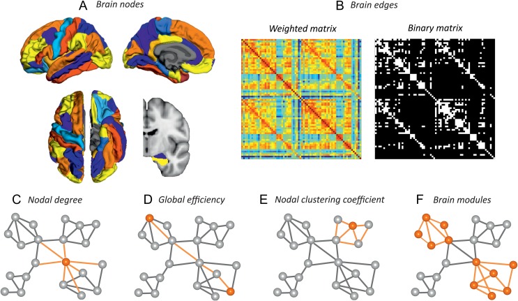
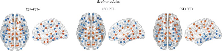
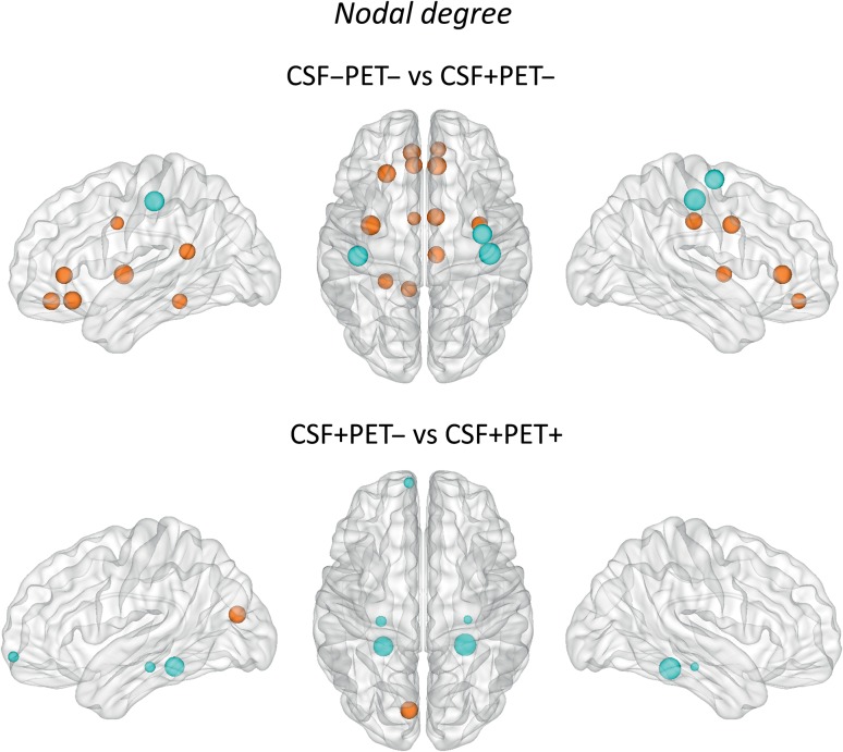
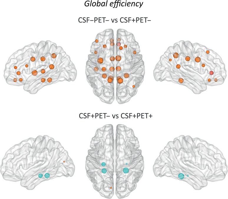
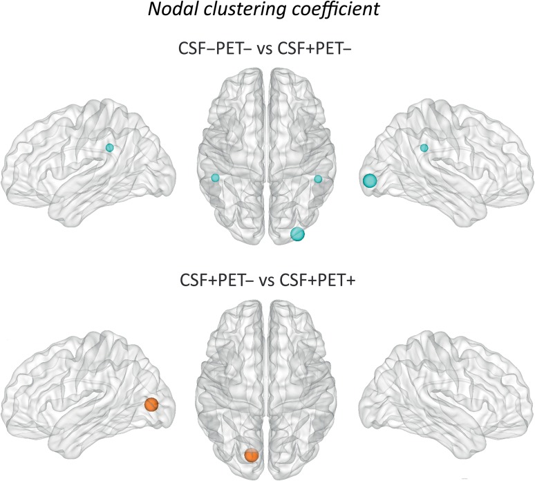
There is increasing evidence showing that the accumulation of the amyloid-β (Aβ) peptide into extracellular plaques is a central event in Alzheimer's disease (AD). These abnormalities can be detected as lowered levels of Aβ42 in the cerebrospinal fluid (CSF) and are followed by increased amyloid burden on positron emission tomography (PET) several years before the onset of dementia. The aim of this study was to assess amyloid network topology in nondemented individuals with early stage Aβ accumulation, defined as abnormal CSF Aβ42 levels and normal Florbetapir PET (CSF+/PET-), and more advanced Aβ accumulation, defined as both abnormal CSF Aβ42 and Florbetapir PET (CSF+/PET+). The amyloid networks were built using correlations in the mean 18F-florbetapir PET values between 72 brain regions and analyzed using graph theory analyses. Our findings showed an association between early amyloid stages and increased covariance as well as shorter paths between several brain areas that overlapped with the default-mode network (DMN). Moreover, we found that individuals with more advanced amyloid accumulation showed more widespread changes in brain regions both within and outside the DMN. These findings suggest that amyloid network topology could potentially be used to assess disease progression in the predementia stages of AD.
越来越多的证据表明,β淀粉样蛋白(Aβ)肽在细胞外斑块中的积累是阿尔茨海默病(AD)的中心事件。这些异常可以通过脑脊液(CSF)中 Aβ42 水平降低来检测,并在痴呆症发作前几年通过正电子发射断层扫描(PET)显示出淀粉样蛋白负担增加。本研究旨在评估早期 Aβ 积累的非痴呆个体的淀粉样蛋白网络拓扑结构,定义为 CSF Aβ42 水平异常和 Florbetapir PET 正常(CSF+/PET-),以及更严重的 Aβ 积累,定义为 CSF Aβ42 和 Florbetapir PET 均异常(CSF+/PET+)。使用 72 个大脑区域之间的平均 18F-Florbetapir PET 值的相关性构建淀粉样蛋白网络,并使用图论分析进行分析。我们的研究结果表明,早期淀粉样蛋白阶段与几个大脑区域之间的协方差增加以及路径缩短有关,这些区域与默认模式网络(DMN)重叠。此外,我们发现,具有更严重淀粉样蛋白积累的个体在 DMN 内外的大脑区域显示出更广泛的变化。这些发现表明,淀粉样蛋白网络拓扑结构可能可用于评估 AD 痴呆前阶段的疾病进展。