Department of Diagnostic, Clinic and Public Health Medicine, University of Modena and Reggio Emilia, Via del Pozzo 87, 41125, Modena, Italy.
Iranian Research Center for HIV/AIDS, Tehran University of Medical Sciences, Tehran, Iran.
Ann Clin Microbiol Antimicrob. 2017 Nov 14;16(1):72. doi: 10.1186/s12941-017-0246-5.
Recently, we published data suggesting a mutualistic relationship between HSV-1 and Candida. albicans; in particular: (a) HSV-1 infected macrophages are inhibited in their anti-Candida effector function and (b) Candida biofilm protects HSV-1 from inactivation. The present in vitro study is aimed at testing the effects of Candida biofilm on HSV-1 sensitivity to pharmacological and physical stress, such as antiviral drugs (acyclovir and foscarnet) and laser UVA1 irradiation. We also investigated whether fungus growth pattern, either sessile or planktonic, influences HSV-1 sensitivity to antivirals.
Mature Candida biofilms were exposed to HSV-1 and then irradiated with laser light (UVA1, 355 λ). In another set of experiments, mature Candida biofilm were co-cultured with HSV-1 infected VERO cells in the presence of different concentrations of acyclovir or foscarnet. In both protocols, controls unexposed to laser or drugs were included. The viral yield of treated and untreated samples was evaluated by end-point titration. To evaluate whether this protective effect might occur in relation with a different growth pattern, HSV-1 infected cells were co-cultured with either sessile or planktonic forms of Candida and then assessed for susceptibility to antiviral drugs.
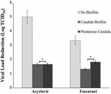
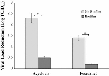
UVA1 irradiation caused a 2 Log reduction of virus yield in the control cultures whereas the reduction was only 1 Log with Candida biofilm, regardless to the laser dose applied to the experimental samples (50 or 100 J/cm). The presence of biofilm increased the IC from 18.4-25.6 J/cm. Acyclovir caused a 2.3 Log reduction of virus yield in the control cultures whereas with Candida biofilm the reduction was only 0.5 Log; foscarnet determined a reduction of 1.4 Log in the controls and 0.2 Log in biofilm cultures. Consequently, the ICs for acyclovir and foscarnet increased by 4- and 12-folds, respectively, compared to controls. When HSV-1 was exposed to either sessile or planktonic fungal cells, the antiviral treatments caused approximately the same weak reduction of virus yield.
These data demonstrate that: (1) HSV-1 encompassed in Candida biofilm is protected from inactivation by physical (laser) and pharmacological (acyclovir or foscarnet) treatments; (2) the drug antiviral activity is reduced at a similar extent for both sessile or planktonic Candida.
最近,我们发表的数据表明单纯疱疹病毒 1(HSV-1)和白色念珠菌之间存在共生关系;具体来说:(a)HSV-1 感染的巨噬细胞在抗白色念珠菌效应功能中受到抑制;(b)白色念珠菌生物膜保护 HSV-1 免受失活。本体外研究旨在测试白色念珠菌生物膜对 HSV-1 对药物和物理应激(如抗病毒药物(阿昔洛韦和膦甲酸)和激光 UVA1 照射)敏感性的影响。我们还研究了真菌生长模式(静止或浮游)是否会影响 HSV-1 对抗病毒药物的敏感性。
成熟的白色念珠菌生物膜暴露于 HSV-1 后,用激光(UVA1,355λ)照射。在另一组实验中,成熟的白色念珠菌生物膜与 HSV-1 感染的 VERO 细胞在不同浓度的阿昔洛韦或膦甲酸存在下共培养。在这两个方案中,都包括未暴露于激光或药物的对照。通过终点滴定法评估处理和未处理样品的病毒产量。为了评估这种保护作用是否与不同的生长模式有关,将感染 HSV-1 的细胞与白色念珠菌的静止或浮游形式共培养,然后评估其对抗病毒药物的敏感性。
UVA1 照射导致对照培养物中的病毒产量降低 2 个对数,但无论应用于实验样品的激光剂量(50 或 100 J/cm)如何,生物膜的存在仅降低了 1 个对数。生物膜的存在使 IC 从 18.4-25.6 J/cm 增加。阿昔洛韦使对照培养物中的病毒产量降低了 2.3 个对数,而生物膜中的降低仅为 0.5 个对数;膦甲酸在对照培养物中降低了 1.4 个对数,在生物膜培养物中降低了 0.2 个对数。因此,与对照相比,阿昔洛韦和膦甲酸的 IC 分别增加了 4 倍和 12 倍。当 HSV-1 暴露于静止或浮游真菌细胞时,抗病毒治疗导致病毒产量的弱降低程度大致相同。
这些数据表明:(1)被白色念珠菌生物膜包裹的 HSV-1 受到物理(激光)和药物(阿昔洛韦或膦甲酸)处理的失活保护;(2)药物抗病毒活性对静止或浮游白色念珠菌的降低程度大致相同。