Wang Yanping, Xu Congying, Zhai Liping, Lu Xudong, Wu Xiaoqiang, Yi Yahui, Liu Ziyun, Guan Qiaobing, Zhang Xiaoling
Department of Neurology, the Second Hospital of Jiaxing City, Jiaxing, Zhejiang.
Department of Radiology, the Second Hospital of Jiaxing City, Jiaxing, Zhejiang, China.
J Pain Res. 2017 Dec 5;10:2741-2750. doi: 10.2147/JPR.S143734. eCollection 2017.
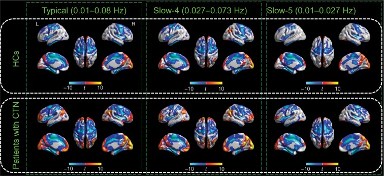
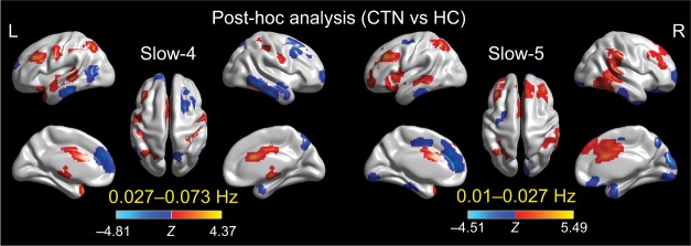
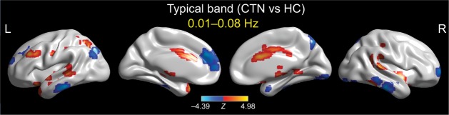
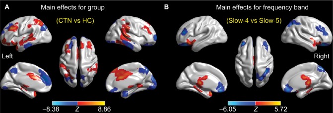
Resting-state functional magnetic resonance imaging (R-fMRI) signals are spatiotemporally organized. R-fMRI studies in patients with classic trigeminal neuralgia (CTN) have suggested alterations in functional connectivity. However, far less attention has been given to investigations of the local oscillations and their frequency-specific changes in these patients. The objective of this study was to address this issue in patients with CTN. R-fMRI data from 17 patients with CTN and 19 age- and gender-matched healthy controls (HCs) were analyzed using amplitude of low-frequency fluctuation (ALFF). The ALFF was computed across different frequencies (slow-4: 0.027-0.073 Hz; slow-5: 0.01-0.027 Hz; and typical band: 0.01-0.08 Hz) in patients with CTN compared to HCs. In the typical band, patients with CTN showed increases of ALFF in bilateral temporal, occipital, and left middle frontal regions and in the left middle cingulate gyrus, as well as decreases of ALFF in the right inferior temporal region and in regions (medial prefrontal regions) of default mode network. These significant group differences were identified in different sub-bands, with greater brainstem findings in higher frequencies (slow-4) and extensive default mode network and right postparietal results in lower frequencies (slow-5). Furthermore, significant relationships were found between subjective pain ratings and both amplitudes of higher frequency (slow-4) blood oxygen level-dependent (BOLD) signals in pain localization brain regions and lower frequencies (slow-5) in pain signaling/modulating brain regions in the patients, and decreased ALFF within the prefrontal regions was significantly correlated with pain duration in the patients. This result supports our hypothesis that trigeminal pain has a characteristic spatiotemporal distribution of low-frequency BOLD signals. These findings might contribute to a better understanding of the impact of CTN on the brain's intrinsic architecture. Future studies should take the frequencies into account when measuring brain resting BOLD signals of patients with CTN.
静息态功能磁共振成像(R-fMRI)信号在时空上是有组织的。对经典三叉神经痛(CTN)患者的R-fMRI研究表明其功能连接存在改变。然而,这些患者局部振荡及其频率特异性变化的研究却很少受到关注。本研究的目的是解决CTN患者中的这一问题。使用低频波动幅度(ALFF)分析了17例CTN患者和19例年龄及性别匹配的健康对照(HCs)的R-fMRI数据。与HCs相比,计算了CTN患者不同频率(慢4:0.027 - 0.073Hz;慢5:0.01 - 0.027Hz;以及典型频段:0.01 - 0.08Hz)的ALFF。在典型频段,CTN患者双侧颞叶、枕叶、左侧额中回以及左侧中央扣带回的ALFF增加,而右侧颞下回以及默认模式网络区域(内侧前额叶区域)的ALFF降低。这些显著的组间差异在不同子频段中被识别出来,在较高频率(慢4)时脑干发现更为明显,而在较低频率(慢5)时默认模式网络和右侧顶叶后部的结果更为广泛。此外,在患者中发现主观疼痛评分与疼痛定位脑区高频(慢4)血氧水平依赖(BOLD)信号的幅度以及疼痛信号/调节脑区低频(慢5)的幅度均存在显著关系,并且前额叶区域内ALFF的降低与患者的疼痛持续时间显著相关。这一结果支持了我们的假设,即三叉神经痛具有低频BOLD信号的特征性时空分布。这些发现可能有助于更好地理解CTN对大脑固有结构的影响。未来在测量CTN患者的脑静息BOLD信号时应考虑频率因素。