• 文献检索
  • 文档翻译
  • 深度研究
  • 学术资讯
  • Suppr Zotero 插件Zotero 插件
  • 邀请有礼
  • 套餐&价格
  • 历史记录
应用&插件
Suppr Zotero 插件Zotero 插件浏览器插件Mac 客户端Windows 客户端微信小程序
定价
高级版会员购买积分包购买API积分包
服务
文献检索文档翻译深度研究API 文档MCP 服务
关于我们
关于 Suppr公司介绍联系我们用户协议隐私条款
关注我们

Suppr 超能文献

核心技术专利:CN118964589B侵权必究
粤ICP备2023148730 号-1Suppr @ 2026

文献检索

告别复杂PubMed语法,用中文像聊天一样搜索,搜遍4000万医学文献。AI智能推荐,让科研检索更轻松。

立即免费搜索

文件翻译

保留排版,准确专业,支持PDF/Word/PPT等文件格式,支持 12+语言互译。

免费翻译文档

深度研究

AI帮你快速写综述,25分钟生成高质量综述,智能提取关键信息,辅助科研写作。

立即免费体验

秀丽隐杆线虫神经胶质类似物 glit-1 中的突变、凋亡途径和钙伴侣 crt-1 增加了 6-OHDA 处理后的多巴胺能神经退行性变。

Mutations in Caenorhabditis elegans neuroligin-like glit-1, the apoptosis pathway and the calcium chaperone crt-1 increase dopaminergic neurodegeneration after 6-OHDA treatment.

机构信息

School of Life Sciences, University of Dundee, Dundee, United Kingdom.

出版信息

PLoS Genet. 2018 Jan 18;14(1):e1007106. doi: 10.1371/journal.pgen.1007106. eCollection 2018 Jan.

DOI:10.1371/journal.pgen.1007106
PMID:29346364
原文链接:https://pmc.ncbi.nlm.nih.gov/articles/PMC5773152/
Abstract

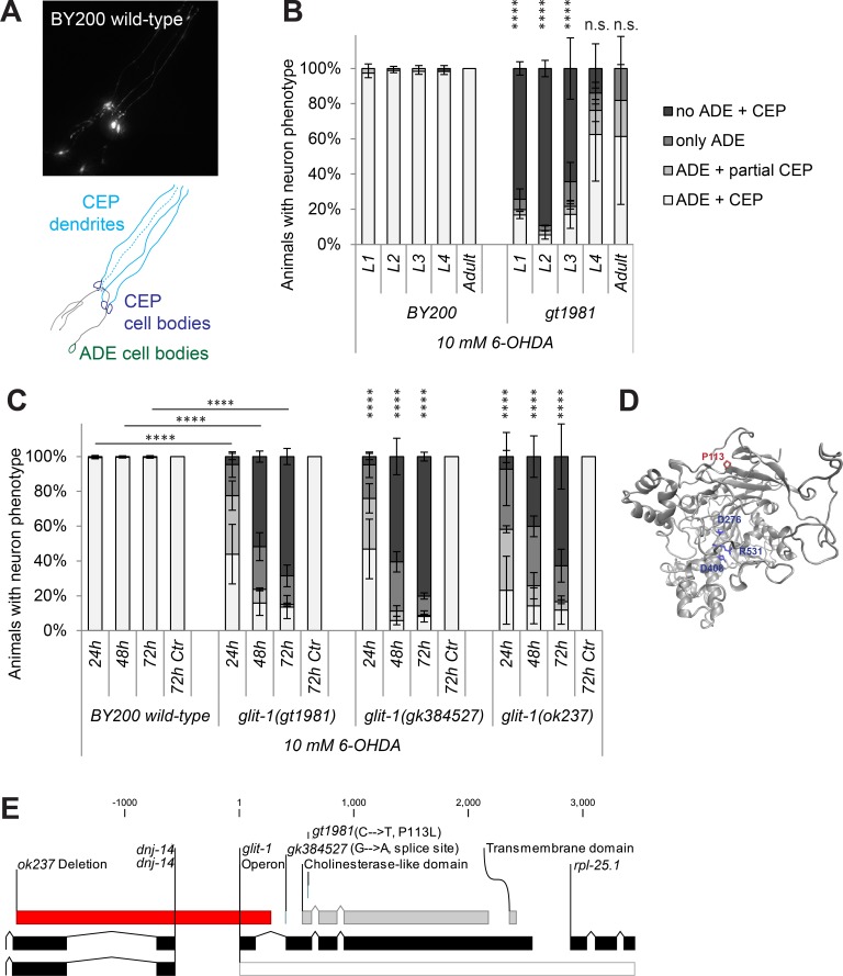
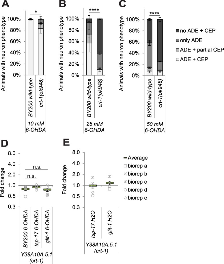
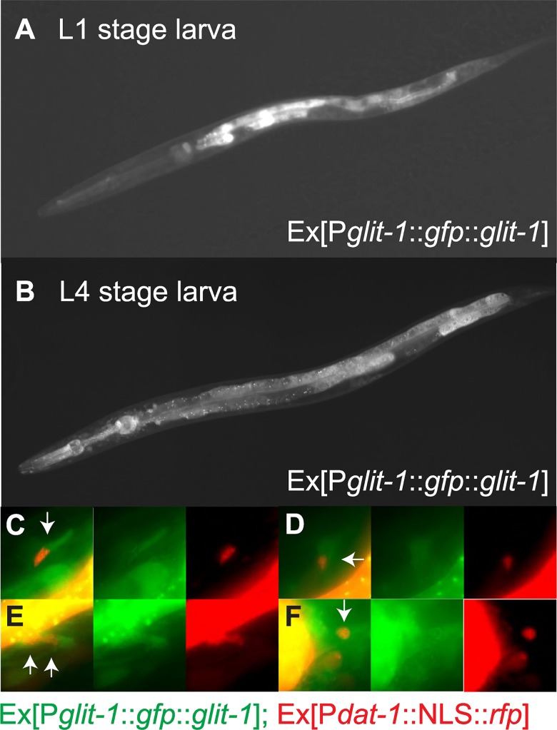
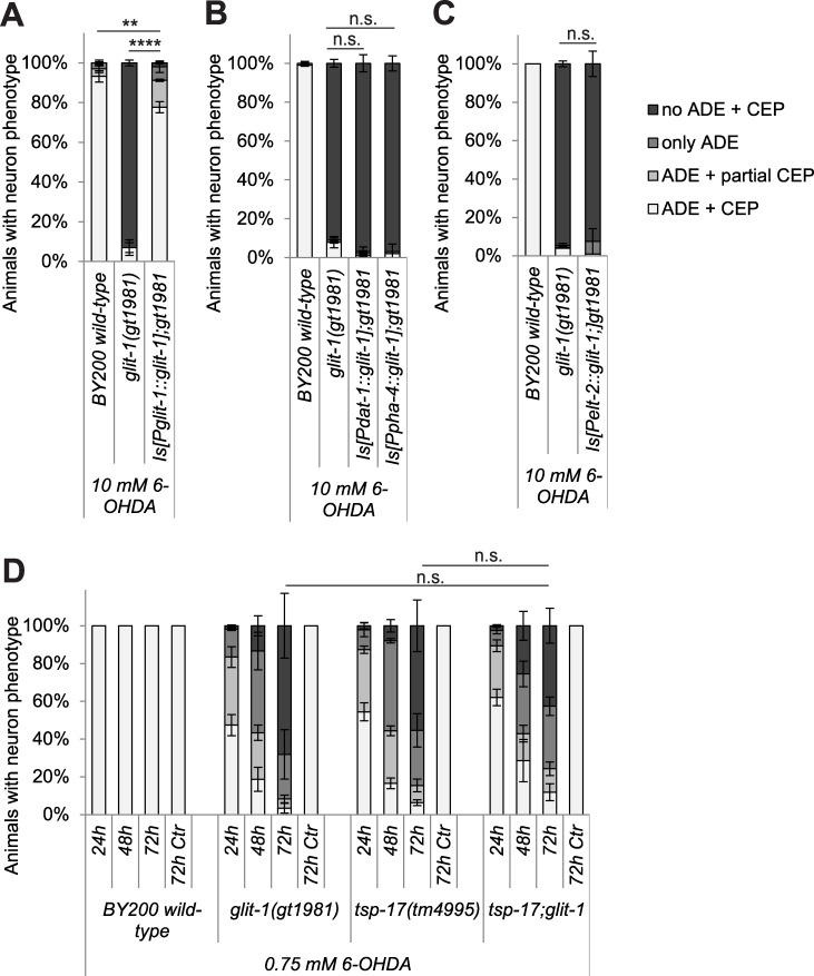
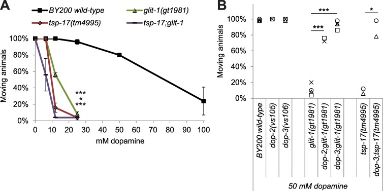
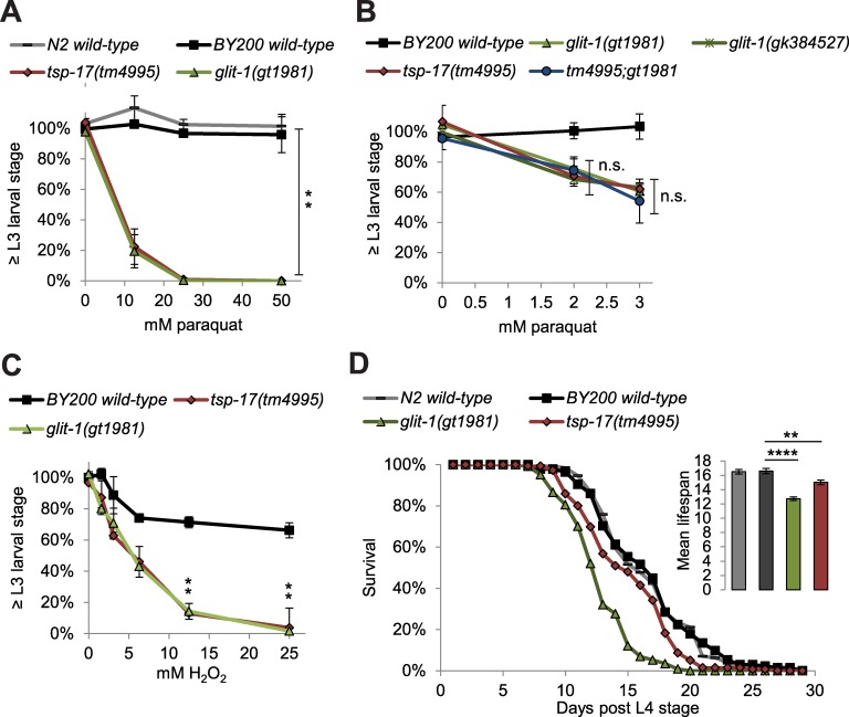
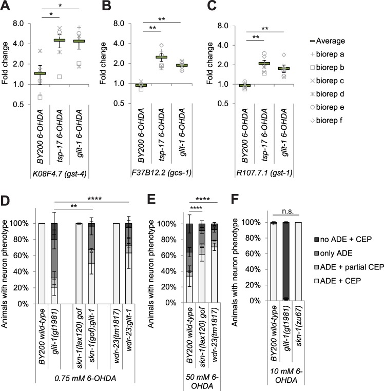
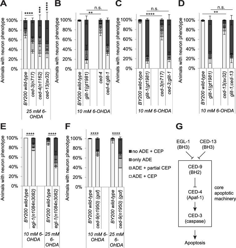
The loss of dopaminergic neurons is a hallmark of Parkinson's disease, the aetiology of which is associated with increased levels of oxidative stress. We used C. elegans to screen for genes that protect dopaminergic neurons against oxidative stress and isolated glit-1 (gliotactin (Drosophila neuroligin-like) homologue). Loss of the C. elegans neuroligin-like glit-1 causes increased dopaminergic neurodegeneration after treatment with 6-hydroxydopamine (6-OHDA), an oxidative-stress inducing drug that is specifically taken up into dopaminergic neurons. Furthermore, glit-1 mutants exhibit increased sensitivity to oxidative stress induced by H2O2 and paraquat. We provide evidence that GLIT-1 acts in the same genetic pathway as the previously identified tetraspanin TSP-17. After exposure to 6-OHDA and paraquat, glit-1 and tsp-17 mutants show almost identical, non-additive hypersensitivity phenotypes and exhibit highly increased induction of oxidative stress reporters. TSP-17 and GLIT-1 are both expressed in dopaminergic neurons. In addition, the neuroligin-like GLIT-1 is expressed in pharynx, intestine and several unidentified cells in the head. GLIT-1 is homologous, but not orthologous to neuroligins, transmembrane proteins required for the function of synapses. The Drosophila GLIT-1 homologue Gliotactin in contrast is required for epithelial junction formation. We report that GLIT-1 likely acts in multiple tissues to protect against 6-OHDA, and that the epithelial barrier of C. elegans glit-1 mutants does not appear to be compromised. We further describe that hyperactivation of the SKN-1 oxidative stress response pathway alleviates 6-OHDA-induced neurodegeneration. In addition, we find that mutations in the canonical apoptosis pathway and the calcium chaperone crt-1 cause increased 6-OHDA-induced dopaminergic neuron loss. In summary, we report that the neuroligin-like GLIT-1, the canonical apoptosis pathway and the calreticulin CRT-1 are required to prevent 6-OHDA-induced dopaminergic neurodegeneration.

摘要

多巴胺能神经元的丧失是帕金森病的一个标志,其病因与氧化应激水平升高有关。我们使用秀丽隐杆线虫筛选出能保护多巴胺能神经元免受氧化应激的基因,并分离出 glit-1(神经胶质蛋白(果蝇神经连接蛋白样)同源物)。秀丽隐杆线虫神经连接蛋白样 glit-1 的缺失会导致在 6-羟基多巴胺(一种专门进入多巴胺能神经元的氧化应激诱导药物)处理后多巴胺能神经退行性变增加。此外,glit-1 突变体对 H2O2 和百草枯诱导的氧化应激更敏感。我们提供的证据表明,GLIT-1 与先前鉴定的四跨膜蛋白 TSP-17 处于相同的遗传途径中。在暴露于 6-OHDA 和百草枯后,glit-1 和 tsp-17 突变体表现出几乎相同的、非累加性的超敏表型,并表现出高度增加的氧化应激报告基因诱导。TSP-17 和 GLIT-1 均在多巴胺能神经元中表达。此外,神经连接蛋白样 GLIT-1 还在咽、肠和头部的几个未识别细胞中表达。GLIT-1 与神经连接蛋白同源,但不是同系物,神经连接蛋白是突触功能所必需的跨膜蛋白。相比之下,果蝇 GLIT-1 同源物 Gliotactin 对于上皮连接形成是必需的。我们报告说,GLIT-1 可能在多种组织中发挥作用以抵抗 6-OHDA,并且 C. elegans glit-1 突变体的上皮屏障似乎没有受到损害。我们进一步描述说,SKN-1 氧化应激反应途径的过度激活可减轻 6-OHDA 诱导的神经退行性变。此外,我们发现经典凋亡途径和钙伴侣 CRT-1 的突变会导致 6-OHDA 诱导的多巴胺能神经元丢失增加。总之,我们报告说神经连接蛋白样 GLIT-1、经典凋亡途径和钙结合蛋白 CRT-1 对于预防 6-OHDA 诱导的多巴胺能神经退行性变是必需的。

https://cdn.ncbi.nlm.nih.gov/pmc/blobs/26fd/5773152/2a55d70776a9/pgen.1007106.g008.jpg
https://cdn.ncbi.nlm.nih.gov/pmc/blobs/26fd/5773152/946fa3cbc291/pgen.1007106.g001.jpg
https://cdn.ncbi.nlm.nih.gov/pmc/blobs/26fd/5773152/40b7f2dc9ad4/pgen.1007106.g002.jpg
https://cdn.ncbi.nlm.nih.gov/pmc/blobs/26fd/5773152/8044c7ecf682/pgen.1007106.g003.jpg
https://cdn.ncbi.nlm.nih.gov/pmc/blobs/26fd/5773152/2be24524c468/pgen.1007106.g004.jpg
https://cdn.ncbi.nlm.nih.gov/pmc/blobs/26fd/5773152/fcb64a4f4e9c/pgen.1007106.g005.jpg
https://cdn.ncbi.nlm.nih.gov/pmc/blobs/26fd/5773152/3cd504202c88/pgen.1007106.g006.jpg
https://cdn.ncbi.nlm.nih.gov/pmc/blobs/26fd/5773152/d481aba8f67d/pgen.1007106.g007.jpg
https://cdn.ncbi.nlm.nih.gov/pmc/blobs/26fd/5773152/2a55d70776a9/pgen.1007106.g008.jpg

相似文献

1
Mutations in Caenorhabditis elegans neuroligin-like glit-1, the apoptosis pathway and the calcium chaperone crt-1 increase dopaminergic neurodegeneration after 6-OHDA treatment.秀丽隐杆线虫神经胶质类似物 glit-1 中的突变、凋亡途径和钙伴侣 crt-1 增加了 6-OHDA 处理后的多巴胺能神经退行性变。
PLoS Genet. 2018 Jan 18;14(1):e1007106. doi: 10.1371/journal.pgen.1007106. eCollection 2018 Jan.
2
6-OHDA-induced dopaminergic neurodegeneration in Caenorhabditis elegans is promoted by the engulfment pathway and inhibited by the transthyretin-related protein TTR-33.6-OHDA 诱导的秀丽隐杆线虫多巴胺能神经退行性变是由吞噬途径促进的,而转甲状腺素相关蛋白 TTR-33 抑制。
PLoS Genet. 2018 Jan 18;14(1):e1007125. doi: 10.1371/journal.pgen.1007125. eCollection 2018 Jan.
3
Tetraspanin (TSP-17) protects dopaminergic neurons against 6-OHDA-induced neurodegeneration in C. elegans.四跨膜蛋白(TSP-17)可保护多巴胺能神经元免受6-羟基多巴胺诱导的秀丽隐杆线虫神经退行性变的影响。
PLoS Genet. 2014 Dec 4;10(12):e1004767. doi: 10.1371/journal.pgen.1004767. eCollection 2014 Dec.
4
Dysregulated LRRK2 signaling in response to endoplasmic reticulum stress leads to dopaminergic neuron degeneration in C. elegans.LRRK2 信号失调对内质网应激的反应导致线虫中多巴胺能神经元的退化。
PLoS One. 2011;6(8):e22354. doi: 10.1371/journal.pone.0022354. Epub 2011 Aug 3.
5
Torsin-mediated protection from cellular stress in the dopaminergic neurons of Caenorhabditis elegans.托辛蛋白介导的对秀丽隐杆线虫多巴胺能神经元细胞应激的保护作用。
J Neurosci. 2005 Apr 13;25(15):3801-12. doi: 10.1523/JNEUROSCI.5157-04.2005.
6
Chronic high-sugar diet in adulthood protects Caenorhabditis elegans from 6-OHDA-induced dopaminergic neurodegeneration.成年期慢性高糖饮食可保护秀丽隐杆线虫免受 6-OHDA 诱导的多巴胺能神经退行性变。
BMC Biol. 2023 Nov 10;21(1):252. doi: 10.1186/s12915-023-01733-9.
7
Snake Venom-Inspired Novel Peptides Protect against Paraquat-Induced Parkinson's Pathology.受蛇毒启发的新型肽可预防百草枯诱导的帕金森病病理变化。
ACS Chem Neurosci. 2025 Apr 2;16(7):1275-1296. doi: 10.1021/acschemneuro.4c00576. Epub 2025 Mar 17.
8
Polysaccharide Suppresses 6-Hydroxydopamine-Induced Neurotoxicity in .多糖抑制6-羟基多巴胺诱导的神经毒性。 (你提供的原文不完整,“in”后面缺少具体内容)
Oxid Med Cell Longev. 2016;2016:4856761. doi: 10.1155/2016/4856761. Epub 2016 Nov 3.
9
Neurogrit Gold Attenuates 6-OHDA-Induced Dopaminergic Neurodegeneration in Parkinson's Model of Caenorhabditis elegans by Reducing α-Synuclein Accumulation and Pink/Pdr-1 Driven Mitochondrial Dysfunction.神经坚毅金通过减少α-突触核蛋白积累以及Pink/Pdr-1驱动的线粒体功能障碍,减轻秀丽隐杆线虫帕金森模型中6-羟基多巴胺诱导的多巴胺能神经退行性变。
CNS Neurosci Ther. 2025 May;31(5):e70401. doi: 10.1111/cns.70401.
10
6-hydroxydopamine (6-OHDA) Oxidative Stress Assay for Observing Dopaminergic Neuron Loss in .用于观察[具体部位]中多巴胺能神经元损失的6-羟基多巴胺(6-OHDA)氧化应激测定
Bio Protoc. 2018 Sep 20;8(18). doi: 10.21769/BioProtoc.3025.

引用本文的文献

1
Unraveling Molecular Targets for Neurodegenerative Diseases Through Models.通过模型揭示神经退行性疾病的分子靶点。
Int J Mol Sci. 2025 Mar 26;26(7):3030. doi: 10.3390/ijms26073030.
2
Tissue distribution of cysteine string protein/DNAJC5 in C. elegans analysed by CRISPR/Cas9-mediated tagging of endogenous DNJ-14.利用 CRISPR/Cas9 介导的内源性 DNJ-14 标记分析秀丽隐杆线虫中半胱氨酸拉链蛋白/DNAJC5 的组织分布。
Cell Tissue Res. 2024 Apr;396(1):41-55. doi: 10.1007/s00441-024-03875-w. Epub 2024 Feb 26.
3
Exploring Caenorhabditis elegans as Parkinson's Disease Model: Neurotoxins and Genetic Implications.

本文引用的文献

1
Oxidative Stress.氧化应激。
Annu Rev Biochem. 2017 Jun 20;86:715-748. doi: 10.1146/annurev-biochem-061516-045037. Epub 2017 Apr 24.
2
Epidemiology, environmental risk factors and genetics of Parkinson's disease.帕金森病的流行病学、环境危险因素及遗传学
Presse Med. 2017 Mar;46(2 Pt 1):175-181. doi: 10.1016/j.lpm.2017.01.001. Epub 2017 Feb 8.
3
Intestinal Autophagy Improves Healthspan and Longevity in C. elegans during Dietary Restriction.肠道自噬在饮食限制期间改善秀丽隐杆线虫的健康寿命和寿命。
探索秀丽隐杆线虫作为帕金森病模型:神经毒素和遗传影响。
Neurotox Res. 2024 Feb 6;42(1):11. doi: 10.1007/s12640-024-00686-3.
4
A Caenorhabditis elegans model of autosomal dominant adult-onset neuronal ceroid lipofuscinosis identifies ethosuximide as a potential therapeutic.秀丽隐杆线虫常染色体显性遗传成人发病神经元蜡样脂褐质沉积症模型鉴定乙琥胺为一种潜在的治疗药物。
Hum Mol Genet. 2023 May 18;32(11):1772-1785. doi: 10.1093/hmg/ddac263.
5
Nanostars Carrying Multifunctional Neurotrophic Dendrimers Protect Neurons in Preclinical In Vitro Models of Neurodegenerative Disorders.纳米星携带多功能神经营养树突状聚合物在神经退行性疾病的临床前体外模型中保护神经元。
ACS Appl Mater Interfaces. 2022 Oct 26;14(42):47445-47460. doi: 10.1021/acsami.2c14220. Epub 2022 Oct 11.
6
Structure and evolution of neuronal wiring receptors and ligands.神经元连接受体和配体的结构与演化。
Dev Dyn. 2023 Jan;252(1):27-60. doi: 10.1002/dvdy.512. Epub 2022 Jul 6.
7
Noncanonical necrosis in 2 different cell types in a Caenorhabditis elegans NAD+ salvage pathway mutant.秀丽隐杆线虫烟酰胺腺嘌呤二核苷酸(NAD+)补救途径突变体中 2 种不同细胞类型的非典型性细胞坏死。
G3 (Bethesda). 2022 Apr 4;12(4). doi: 10.1093/g3journal/jkac033.
8
Modeling Parkinson's Disease: Not Only Rodents?帕金森病建模:不仅仅是啮齿动物?
Front Aging Neurosci. 2021 Aug 6;13:695718. doi: 10.3389/fnagi.2021.695718. eCollection 2021.
9
The Role of Ca Signaling in Aging and Neurodegeneration: Insights from Models.钙信号在衰老和神经退行性变中的作用:模型研究的启示。
Cells. 2020 Jan 14;9(1):204. doi: 10.3390/cells9010204.
10
Control of neural development and function by glial neuroligins.神经胶质 neuroligins 对神经发育和功能的控制。
Curr Opin Neurobiol. 2019 Aug;57:163-170. doi: 10.1016/j.conb.2019.03.007. Epub 2019 Apr 13.
PLoS Genet. 2016 Jul 14;12(7):e1006135. doi: 10.1371/journal.pgen.1006135. eCollection 2016 Jul.
4
Understanding Dopaminergic Cell Death Pathways in Parkinson Disease.了解帕金森病中的多巴胺能细胞死亡途径。
Neuron. 2016 May 18;90(4):675-91. doi: 10.1016/j.neuron.2016.03.038.
5
Programmed cell death and clearance of cell corpses in Caenorhabditis elegans.秀丽隐杆线虫中的程序性细胞死亡与细胞尸体清除
Cell Mol Life Sci. 2016 Jun;73(11-12):2221-36. doi: 10.1007/s00018-016-2196-z. Epub 2016 Apr 5.
6
Two phases of aging separated by the Smurf transition as a public path to death.衰老的两个阶段由蓝精灵转变分隔开来,这是通向死亡的一条共同路径。
Sci Rep. 2016 Mar 22;6:23523. doi: 10.1038/srep23523.
7
Context Specificity of Stress-activated Mitogen-activated Protein (MAP) Kinase Signaling: The Story as Told by Caenorhabditis elegans.应激激活的丝裂原活化蛋白(MAP)激酶信号传导的背景特异性:秀丽隐杆线虫讲述的故事
J Biol Chem. 2016 Apr 8;291(15):7796-804. doi: 10.1074/jbc.R115.711101. Epub 2016 Feb 23.
8
The cellular and molecular landscape of neuroligins.神经连接蛋白的细胞和分子格局
Trends Neurosci. 2015 Aug;38(8):496-505. doi: 10.1016/j.tins.2015.06.004. Epub 2015 Jul 21.
9
Neuronal responses to stress and injury in C. elegans.秀丽隐杆线虫中神经元对应激和损伤的反应。
FEBS Lett. 2015 Jun 22;589(14):1644-52. doi: 10.1016/j.febslet.2015.05.005. Epub 2015 May 13.
10
Tetraspanin (TSP-17) protects dopaminergic neurons against 6-OHDA-induced neurodegeneration in C. elegans.四跨膜蛋白(TSP-17)可保护多巴胺能神经元免受6-羟基多巴胺诱导的秀丽隐杆线虫神经退行性变的影响。
PLoS Genet. 2014 Dec 4;10(12):e1004767. doi: 10.1371/journal.pgen.1004767. eCollection 2014 Dec.