General Surgery Department, Second Xiangya Hospital, Central South University, Changsha, Hunan, 410011, People's Republic of China.
Research Laboratory of Hepatobiliary Diseases, Second Xiangya Hospital, Central South University, Changsha, Hunan, 410011, People's Republic of China.
World J Surg Oncol. 2018 Jan 18;16(1):11. doi: 10.1186/s12957-018-1316-7.
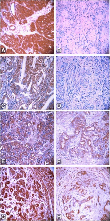
This study investigated UGP2 (uridine diphosphate-glucose pyrophosphorylase-2) and CFL1 (cofilin-1) expression in pancreatic ductal carcinoma (PDC), paracancerous tissue (PT), benign lesions (BL), and normal tissue (NT) and their clinicopathological significance.
Surgical specimens, which were collected from 106 cases of pancreatic ductal carcinoma, 35 cases of paracancerous tissues, 55 cases of benign lesions and 13 cases of normal pancreatic tissues, were fixed with 4% formaldehyde to prepare conventional paraffin-embedded sections. EnVision immunohistochemical was used to stain for UGP2 and CFL1. Kaplan-Meier survival analysis was performed to assess the correlation of expression pattern with survival.
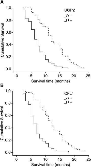
We found that positive UGP2 and CFL1 expression in PDC were significantly higher than those in PT, BL, and NT. In PT and BL with positive UGP2 and CFL1 expression, mild to severe atypical hyperplasia or intraepithelial neoplasia of grades II-III was observed in ductal epithelium. Positive UGP2 and CFL1 expression in cases with high differentiation, no lymph node metastasis, no surrounding invasion, and TNM (tumor-node-metastasis) staging I or/and II were significantly lower than those in cases with poor differentiation, lymph node metastasis, surrounding invasion, and TNM stage III and/or IV. Positive UGP2 expression in male patients was significantly lower than that in female patients. UGP2 and CFL1 expression in PDC were positively correlated. Kaplan-Meier survival analysis showed the degree of differentiation, tumor maximal diameter, TNM stage, lymph node metastasis, and surrounding invasion, and UGP2 and CFL1 expression were closely related to the average survival time of patients with PDC. The survival time of patients with positive UGP2 and CFL1 expression was significantly shorter than that of patients with negative expression. Cox multivariate analysis showed that poor differentiation, tumor maximal diameter ≥ 3 cm, TNM stage III or IV, lymph node metastasis, surrounding invasion, and positive UGP2 and CFL1 expression was negatively correlated with the postoperative survival rate and positively correlated with the mortality of patients with PDC.
Positive expression of UGP2 and CFL1 can serve a valuable prognostic factor in pancreatic cancer.
本研究探讨了 UGP2(尿苷二磷酸葡萄糖焦磷酸化酶-2)和 CFL1(丝切蛋白-1)在胰腺导管腺癌(PDC)、癌旁组织(PT)、良性病变(BL)和正常组织(NT)中的表达及其临床病理意义。
收集 106 例胰腺导管腺癌、35 例癌旁组织、55 例良性病变和 13 例正常胰腺组织的手术标本,用 4%甲醛固定,制备常规石蜡包埋切片。采用 EnVision 免疫组化法检测 UGP2 和 CFL1 的表达。采用 Kaplan-Meier 生存分析评估表达模式与生存的相关性。
我们发现,PDC 中 UGP2 和 CFL1 的阳性表达明显高于 PT、BL 和 NT。在 UGP2 和 CFL1 阳性的 PT 和 BL 中,观察到导管上皮的轻至重度非典型增生或 II-III 级上皮内瘤变。高分化、无淋巴结转移、无周围侵犯、TNM(肿瘤-淋巴结-转移)分期 I 期和/或 II 期患者的 UGP2 和 CFL1 阳性表达明显低于低分化、淋巴结转移、周围侵犯、TNM 分期 III 期和/或 IV 期患者。男性患者的 UGP2 阳性表达明显低于女性患者。PDC 中 UGP2 和 CFL1 的表达呈正相关。Kaplan-Meier 生存分析显示,分化程度、肿瘤最大直径、TNM 分期、淋巴结转移、周围侵犯以及 UGP2 和 CFL1 的表达与 PDC 患者的平均生存时间密切相关。UGP2 和 CFL1 阳性表达患者的生存时间明显短于阴性表达患者。Cox 多因素分析显示,低分化、肿瘤最大直径≥3cm、TNM 分期 III 或 IV 期、淋巴结转移、周围侵犯以及 UGP2 和 CFL1 阳性表达与 PDC 患者术后生存率呈负相关,与死亡率呈正相关。
UGP2 和 CFL1 的阳性表达可作为胰腺癌有价值的预后因素。